Category:Flow cytometry
Jump to navigation
Jump to search
technique of suspending cells in a stream of fluid and passing them by an electronic detection apparatus | |||||
| Upload media | |||||
| Subclass of |
| ||||
|---|---|---|---|---|---|
| |||||
English: Flow cytometry is a method in cell biology that employs the deflection of laser light a well as the excitation of fluorescent dyes to analyse various properties of a high number cells in a relatively short time. This category lists methods and tools used in flow cytometry.
Subcategories
This category has the following 4 subcategories, out of 4 total.
C
- Flow cytometers (5 F)
I
- Immunophenotyping (4 F)
Media in category "Flow cytometry"
The following 200 files are in this category, out of 340 total.
(previous page) (next page)-
-
-
-
-
-
-
-
-
A-Novel-Model-of-Intravital-Platelet-Imaging-Using-CD41-ZsGreen1-Transgenic-Rats-pone.0154661.s002.ogv 25 s, 1,024 × 348; 3.32 MB
-
A-Novel-Model-of-Intravital-Platelet-Imaging-Using-CD41-ZsGreen1-Transgenic-Rats-pone.0154661.s003.ogv 25 s, 1,024 × 348; 359 KB
-
-
-
-
-
-
-
A-Plasmodium-falciparum-Strain-Expressing-GFP-throughout-the-Parasite's-Life-Cycle-pone.0009156.s001.ogv 12 s, 1,388 × 1,040; 64 KB
-
A-Plasmodium-falciparum-Strain-Expressing-GFP-throughout-the-Parasite's-Life-Cycle-pone.0009156.s002.ogv 24 s, 1,388 × 1,040; 666 KB
-
-
-
-
An-intraductal-human-in-mouse-transplantation-model-mimics-the-subtypes-of-ductal-carcinoma-in-situ-bcr2358-S1.ogv 5 min 44 s, 320 × 240; 9.76 MB
-
Analysis-of-Cells-Targeted-by-Salmonella-Type-III-Secretion-In-Vivo-ppat.0030196.sv001.ogv 16 s, 558 × 524; 264 KB
-
Analysis-of-Cells-Targeted-by-Salmonella-Type-III-Secretion-In-Vivo-ppat.0030196.sv002.ogv 13 s, 542 × 476; 254 KB
-
Analysis-of-Cells-Targeted-by-Salmonella-Type-III-Secretion-In-Vivo-ppat.0030196.sv003.ogv 13 s, 542 × 476; 175 KB
-
Analysis-of-Cells-Targeted-by-Salmonella-Type-III-Secretion-In-Vivo-ppat.0030196.sv004.ogv 13 s, 542 × 476; 195 KB
-
Analysis-of-Cells-Targeted-by-Salmonella-Type-III-Secretion-In-Vivo-ppat.0030196.sv005.ogv 13 s, 542 × 476; 204 KB
-
-
-
-
-
-
AutomatedFlowPipeline.svg 612 × 459; 238 KB
-
-
-
Biofilm-Induced-Tolerance-towards-Antimicrobial-Peptides-pone.0001891.s001.ogv 0.0 s, 512 × 480; 18.13 MB
-
-
-
-
-
Cell Sorting Diagram.jpg 2,784 × 4,032; 10.04 MB
-
Cells-Expressing-the-CEBPbeta-Isoform-LIP-Engulf-Their-Neighbors-pone.0041807.s006.ogv 23 s, 640 × 480; 655 KB
-
Cells-Expressing-the-CEBPbeta-Isoform-LIP-Engulf-Their-Neighbors-pone.0041807.s007.ogv 53 s, 512 × 500; 1.99 MB
-
Cells-Expressing-the-CEBPbeta-Isoform-LIP-Engulf-Their-Neighbors-pone.0041807.s008.ogv 8.5 s, 1,392 × 1,040; 423 KB
-
Cells-Expressing-the-CEBPbeta-Isoform-LIP-Engulf-Their-Neighbors-pone.0041807.s009.ogv 13 s, 434 × 348; 131 KB
-
Cells-Expressing-the-CEBPbeta-Isoform-LIP-Engulf-Their-Neighbors-pone.0041807.s010.ogv 21 s, 530 × 512; 754 KB
-
Cells-Expressing-the-CEBPbeta-Isoform-LIP-Engulf-Their-Neighbors-pone.0041807.s011.ogv 25 s, 512 × 512; 276 KB
-
-
-
-
-
-
-
-
Circadian-Clocks-in-Mouse-and-Human-CD4+-T-Cells-pone.0029801.s002.ogv 12 s, 256 × 256; 2.15 MB
-
Contribution-of-Caspase(s)-to-the-Cell-Cycle-Regulation-at-Mitotic-Phase-pone.0018449.s002.ogv 24 s, 450 × 376; 318 KB
-
Contribution-of-Caspase(s)-to-the-Cell-Cycle-Regulation-at-Mitotic-Phase-pone.0018449.s003.ogv 24 s, 450 × 376; 426 KB
-
Contribution-of-Caspase(s)-to-the-Cell-Cycle-Regulation-at-Mitotic-Phase-pone.0018449.s004.ogv 24 s, 450 × 376; 470 KB
-
Contribution-of-Caspase(s)-to-the-Cell-Cycle-Regulation-at-Mitotic-Phase-pone.0018449.s005.ogv 24 s, 450 × 376; 428 KB
-
Contribution-of-Caspase(s)-to-the-Cell-Cycle-Regulation-at-Mitotic-Phase-pone.0018449.s006.ogv 24 s, 450 × 376; 477 KB
-
Contribution-of-Caspase(s)-to-the-Cell-Cycle-Regulation-at-Mitotic-Phase-pone.0018449.s007.ogv 49 s, 680 × 512; 2.09 MB
-
Contribution-of-Caspase(s)-to-the-Cell-Cycle-Regulation-at-Mitotic-Phase-pone.0018449.s008.ogv 49 s, 680 × 512; 1.41 MB
-
Crystallization-of-DNA-coated-colloids-ncomms8253-s2.ogv 21 s, 696 × 520; 26.32 MB
-
Crystallization-of-DNA-coated-colloids-ncomms8253-s3.ogv 23 s, 527 × 167; 776 KB
-
Crystallization-of-DNA-coated-colloids-ncomms8253-s4.ogv 18 s, 696 × 520; 15.03 MB
-
Crystallization-of-DNA-coated-colloids-ncomms8253-s5.ogv 15 s, 696 × 520; 20.47 MB
-
Crystallization-of-DNA-coated-colloids-ncomms8253-s6.ogv 17 s, 696 × 520; 23.61 MB
-
Crystallization-of-DNA-coated-colloids-ncomms8253-s7.ogv 17 s, 696 × 520; 27.44 MB
-
Crystallization-of-DNA-coated-colloids-ncomms8253-s8.ogv 20 s, 384 × 288; 11.91 MB
-
Crystallization-of-DNA-coated-colloids-ncomms8253-s9.ogv 20 s, 720 × 576; 14.23 MB
-
-
-
CXCR7-Functions-as-a-Scavenger-for-CXCL12-and-CXCL11-pone.0009175.s001.ogv 6.3 s, 335 × 311; 243 KB
-
CXCR7-Functions-as-a-Scavenger-for-CXCL12-and-CXCL11-pone.0009175.s002.ogv 5.7 s, 321 × 340; 186 KB
-
Cytometer ru.svg 924 × 624; 77 KB
-
Cytometer.svg 924 × 624; 65 KB
-
Cytotoxic-Effect-of-Poly-Dispersed-Single-Walled-Carbon-Nanotubes-on-Erythrocytes-In-Vitro-and-In-pone.0022032.s001.ogv 9.3 s, 1,280 × 1,024; 2.54 MB
-
-
-
-
-
-
Development-of-a-macromolecular-prodrug-for-the-treatment-of-inflammatory-arthritis-mechanisms-ar3130-S1.ogv 8.3 s, 992 × 1,040; 689 KB
-
-
Development-of-an-In-Vitro-Model-for-the-Multi-Parametric-Quantification-of-the-Cellular-pone.0032621.s011.ogv 1 min 10 s, 640 × 480; 11.91 MB
-
-
-
-
-
-
Duchflusszytometer.png 3,160 × 3,035; 476 KB
-
-
-
-
-
-
-
Dynamics-of-Macrophage-Trogocytosis-of-Rituximab-Coated-B-Cells-pone.0014498.s003.ogv 4.0 s, 112 × 148; 48 KB
-
Dynamics-of-Macrophage-Trogocytosis-of-Rituximab-Coated-B-Cells-pone.0014498.s004.ogv 4.0 s, 224 × 572; 265 KB
-
Dynamics-of-Macrophage-Trogocytosis-of-Rituximab-Coated-B-Cells-pone.0014498.s005.ogv 3.9 s, 188 × 164; 233 KB
-
Dynamics-of-Macrophage-Trogocytosis-of-Rituximab-Coated-B-Cells-pone.0014498.s006.ogv 4.0 s, 352 × 208; 236 KB
-
Dynamics-of-Macrophage-Trogocytosis-of-Rituximab-Coated-B-Cells-pone.0014498.s007.ogv 4.0 s, 572 × 320; 336 KB
-
Dynamics-of-Macrophage-Trogocytosis-of-Rituximab-Coated-B-Cells-pone.0014498.s008.ogv 4.0 s, 200 × 500; 257 KB
-
Dynamics-of-Macrophage-Trogocytosis-of-Rituximab-Coated-B-Cells-pone.0014498.s009.ogv 0.7 s, 1,344 × 1,024; 871 KB
-
-
-
-
Entamoeba-histolytica-Cysteine-Proteinase-5-Evokes-Mucin-Exocytosis-from-Colonic-Goblet-Cells-via-ppat.1005579.s004.ogv 4.9 s, 1,104 × 1,104; 6.74 MB
-
Entamoeba-histolytica-Cysteine-Proteinase-5-Evokes-Mucin-Exocytosis-from-Colonic-Goblet-Cells-via-ppat.1005579.s005.ogv 4.5 s, 1,236 × 1,236; 4.43 MB
-
-
-
Extensive-Fusion-of-Mitochondria-in-Spinal-Cord-Motor-Neurons-pone.0038435.s001.ogv 5.8 s, 1,441 × 404; 570 KB
-
Extensive-Fusion-of-Mitochondria-in-Spinal-Cord-Motor-Neurons-pone.0038435.s002.ogv 6.8 s, 982 × 287; 209 KB
-
Extensive-Fusion-of-Mitochondria-in-Spinal-Cord-Motor-Neurons-pone.0038435.s003.ogv 5.9 s, 961 × 207; 134 KB
-
Extensive-Fusion-of-Mitochondria-in-Spinal-Cord-Motor-Neurons-pone.0038435.s004.ogv 18 s, 1,396 × 336; 678 KB
-
Extensive-Fusion-of-Mitochondria-in-Spinal-Cord-Motor-Neurons-pone.0038435.s005.ogv 18 s, 1,396 × 336; 923 KB
-
Extensive-Fusion-of-Mitochondria-in-Spinal-Cord-Motor-Neurons-pone.0038435.s006.ogv 18 s, 1,396 × 336; 1,021 KB
-
-
Externalized-decondensed-neutrophil-chromatin-occludes-pancreatic-ducts-and-drivespancreatitis-ncomms10973-s3.ogv 6.1 s, 1,388 × 1,040; 6.35 MB
-
-
-
Externalized-decondensed-neutrophil-chromatin-occludes-pancreatic-ducts-and-drivespancreatitis-ncomms10973-s6.ogv 7.6 s, 1,920 × 1,080; 3.53 MB
-
-
-
-
Flow Cy.gif 336 × 322; 3 KB
-
Flow cytometer structure.png 865 × 599; 1.98 MB
-
Flow cytometer.png 852 × 615; 2 MB
-
Flow cytometric detection of plasma cells.png 512 × 108; 32 KB
-
Flow cytometric gating by side scatter and CD45, without labels.png 1,451 × 839; 365 KB
-
Flow cytometric gating by side scatter and CD45.png 1,459 × 837; 389 KB
-
Flow cytometric viability by 7-AAD.png 1,047 × 463; 73 KB
-
Flow cytometry 3Dhistogram.png 375 × 256; 99 KB
-
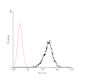
Flow cytometry histogram.png 266 × 268; 4 KB
-
Flow cytometry scatterplot.png 285 × 288; 5 KB
-
Flow cytometry scheme.png 592 × 766; 54 KB
-
Flow-Based-Cytometric-Analysis-of-Cell-Cycle-via-Simulated-Cell-Populations-pcbi.1000741.s007.ogv 50 s, 768 × 576; 2.67 MB
-
FlowCAP Results.png 2,000 × 2,000; 361 KB
-
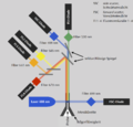
FlowCitometer.svg 607 × 380; 19 KB
-
Flowcytometer MoFlo (DAKO cytomation).jpg 320 × 426; 21 KB
-
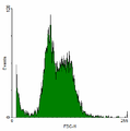
FlowCytometrySFLvsSSC.png 300 × 316; 35 KB
-
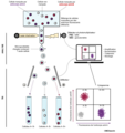
Fluorescence Activated Cell Sorting (FACS) principle.tif 1,201 × 1,280; 380 KB
-
Focalizzazione idrodinamica.png 883 × 750; 21 KB
-
Functional-classification-of-memory-CD8+-T-cells-by-CX3CR1-expression-ncomms9306-s8.ogv 12 s, 640 × 640; 568 KB
-
-
-
-
-
Genetic-epigenesis-pbio.1001325.s014.ogv 3.0 s, 1,548 × 616; 254 KB
-
Genetic-epigenesis-pbio.1001325.s015.ogv 3.0 s, 608 × 302; 48 KB
-
Genetic-epigenesis-pbio.1001325.s016.ogv 4.0 s, 400 × 540; 69 KB
-
Genetic-epigenesis-pbio.1001325.s017.ogv 7.3 s, 1,302 × 626; 187 KB
-
Genetic-epigenesis-pbio.1001325.s018.ogv 7.3 s, 1,302 × 526; 143 KB
-
High-density-lipoprotein-contribute-to-G0-G1S-transition-in-Swiss-NIH3T3-fibroblasts-srep17812-s1.ogv 4.0 s, 236 × 159; 1.13 MB
-
-
-
HIV-AIDS-Immunstatusdiagnostik Kenia.JPG 4,912 × 3,264; 8.75 MB
-
-
IFlow-A-Graphical-User-Interface-for-Flow-Cytometry-Tools-in-Bioconductor-103839.f1.ogv 3 min 16 s, 808 × 624; 4.18 MB
-
-
-
In-Vitro-Formation-of--Cell-Pseudoislets-Using-Islet-Derived-Endothelial-Cells-pone.0072260.s001.ogv 1.4 s, 568 × 576; 364 KB
-
-
-
-
-
-
-
-
-
-
-
-
-
-
-
-
Inside BD LSRFortessa cell analyzer.jpg 4,800 × 3,200; 701 KB
-
-
Lactobacillus-Decelerates-Cervical-Epithelial-Cell-Cycle-Progression-pone.0063592.s003.ogv 19 s, 694 × 520; 3.96 MB
-
Lactobacillus-Decelerates-Cervical-Epithelial-Cell-Cycle-Progression-pone.0063592.s004.ogv 19 s, 694 × 520; 7.11 MB
-
Lactobacillus-Decelerates-Cervical-Epithelial-Cell-Cycle-Progression-pone.0063592.s005.ogv 19 s, 694 × 520; 8.43 MB
-
Lactobacillus-Decelerates-Cervical-Epithelial-Cell-Cycle-Progression-pone.0063592.s006.ogv 19 s, 694 × 520; 3.83 MB
-
Large-scale-cell-production-of-stem-cells-for-clinical-application-using-the-automated-cell-1472-6750-13-102-S4.ogv 3 min 21 s, 400 × 300; 15.43 MB
-
-
-
-
-
-
-
Lineage-Tracking-for-Probing-Heritable-Phenotypes-at-Single-Cell-Resolution-pone.0152395.s008.ogv 30 s, 750 × 393; 2.72 MB
-
-
Long-term-maintenance-of-human-induced-pluripotent-stem-cells-by-automated-cell-culture-system-srep16647-s2.ogv 3 min 31 s, 426 × 240; 4.04 MB
-
-
-
-
-
-
-


