Category:Light microscopy
Jump to navigation
Jump to search
Place images taken through a light microscope in a suitable subcategory of "Category:Light microscopy micrographs". You should not place these images there.
Subcategories
This category has the following 23 subcategories, out of 23 total.
Media in category "Light microscopy"
The following 33 files are in this category, out of 33 total.
-
Acetato-di-sodio 01.jpg 1,200 × 900; 178 KB
-
Braarudosphaera bigelowii 1.webp 251 × 85; 7 KB
-
Chrysochromulina parkeae 1.jpg 700 × 402; 43 KB
-
Chrysochromulina parva 1.jpg 700 × 544; 17 KB
-
Cocobacilos.jpg 900 × 1,600; 107 KB
-
Conchiglia (2).jpg 1,600 × 1,200; 552 KB
-
Conchiglia-a-pois.jpg 1,600 × 1,200; 632 KB
-
Cornea muscle-ad tendon mice cart-horse-surf.png 676 × 650; 847 KB
-
Células de cebolla aumento total 100X en división celular.jpg 654 × 869; 147 KB
-
Dettaglio-conchiglia (1).jpg 1,600 × 1,200; 750 KB
-
Dettaglio-conchiglia (2).jpg 1,600 × 1,200; 710 KB
-
Dettaglio-conchiglia (3).jpg 1,600 × 1,200; 261 KB
-
Droplet Formation in ddPCR.jpg 255 × 240; 11 KB
-
Encapsulation of DNA.jpg 1,920 × 1,440; 220 KB
-
General guide on how to operate an optical microscope with staff from the Stanford Nanofabrication Facility.webm 2 min 8 s, 1,920 × 1,080; 57.55 MB
-
Gold droplets on tungsten coil.jpg 5,423 × 3,187; 1.82 MB
-
HLMVECs.jpg 1,206 × 1,250; 382 KB
-
LRP Sagitt copy.jpg 1,310 × 873; 1.08 MB
-
Marking a microscopy slide.jpg 2,752 × 3,067; 1.93 MB
-
Micrograph of marking a microscopy slide.jpg 4,897 × 2,441; 3.2 MB
-
Microscope Eyepiece Adjustment.jpg 2,400 × 2,400; 427 KB
-
Observation of rocks by Optical microscopy with polarizer.jpg 750 × 686; 158 KB
-
Photograph of marking a microscopy slide.jpg 3,053 × 2,085; 1.43 MB
-
Prymnesiales 1.jpg 1,060 × 940; 79 KB
-
Removing marks from a microscopy slide.jpg 1,961 × 1,037; 520 KB
-
Rhodosporidium toruloides -1588 cells.jpg 2,304 × 4,096; 520 KB
-
Stone1417.png 4,140 × 3,096; 17.95 MB
-
Stone1418.png 4,140 × 3,096; 17.71 MB
-
Stone1476.png 4,140 × 3,096; 16.33 MB
-
Světelná mikroskopie K4H.pdf 1,752 × 1,239; 595 KB
-
Wool-fibres.tif 1,599 × 1,199; 3.72 MB
-
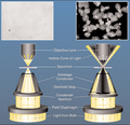
تفاوت نوردهی bright field (سمت چپ) با dark field (سمت راست).png 366 × 355; 156 KB


