Category:Fluorescence microscopy
Jump to navigation
Jump to search
optical microscope that uses fluorescence and phosphorescence | |||||
| Upload media | |||||
| Subclass of | |||||
|---|---|---|---|---|---|
| Part of | |||||
| Named after | |||||
| Has use |
| ||||
| |||||
Subcategories
This category has the following 11 subcategories, out of 11 total.
Media in category "Fluorescence microscopy"
The following 50 files are in this category, out of 50 total.
-
De-Fluoreszenzmikroskopie.ogg 3.4 s; 32 KB
-
0300 Flourescence Stained new.jpg 1,350 × 795; 74 KB
-
0300 Flourescence Stained.jpg 675 × 645; 225 KB
-
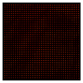
1225 site cold cesium atom array.png 643 × 644; 183 KB
-
13C and 15N incorporation in representative microbial cells.webp 1,752 × 778; 153 KB
-
3D-animation of the diatom Corethron sp.ogv 20 s, 1,240 × 720; 11.7 MB
-
3D-fluorescence imaging for high throughput analysis of microbial eukaryotes.jpg 5,098 × 3,285; 1.39 MB
-
41467 2022 28065 Fig1h-m.jpg 627 × 357; 49 KB
-
41467 2022 28065 Fig1n.jpg 428 × 359; 38 KB
-
41467 2022 28065 Fig1o.jpg 431 × 359; 29 KB
-
7a.png 208 × 211; 38 KB
-
ARC-1034 localization in cells.png 175 × 247; 63 KB
-
Dinophysis tripos.png 2,056 × 2,464; 3.32 MB
-
Encapsulation of DNA.jpg 1,920 × 1,440; 220 KB
-
Filamentous cyanobacteria under confocal fluorescence imaging.jpg 1,949 × 717; 271 KB
-
FLIM-mKO-mCherry.png 996 × 1,107; 682 KB
-
Fluorescence micrograph of Thalassiosira nanae.jpg 1,054 × 1,099; 174 KB
-
Fluorescence microscopy with ZEISS Axiocam (cropped).png 985 × 763; 353 KB
-
Fluorescence microscopy with ZEISS Axiocam.png 1,500 × 767; 588 KB
-
Fluorescence.microscope1.jpg 2,226 × 2,544; 1.09 MB
-
Fluorescence.microscope2.jpg 2,428 × 2,475; 1.21 MB
-
Fluorescence.microscope3.jpg 2,436 × 2,393; 1.26 MB
-
FluorescenceFilters 2008-09-28 cs.svg 836 × 857; 37 KB
-
FluorescenceFilters 2008-09-28-ru.svg 624 × 741; 3 KB
-
FluorescenceFilters 2008-09-28.svg 836 × 857; 37 KB
-
FluorescenceFilters-fr.svg 836 × 857; 26 KB
-
FluorescenceFilters.svg 880 × 745; 82 KB
-
FluorescenceFiltrestraductionFR.jpg 2,000 × 2,050; 203 KB
-
FluorescenceMicroscopeSample HerringSpermSYBRGreen.jpg 2,633 × 1,800; 300 KB
-
Fluorescent green lipid.png 556 × 140; 82 KB
-
Fluoreszenzmikroskopie 2008-09-28.svg 836 × 857; 29 KB
-
Fluoreszenzmikroskopie 2017-03-08.svg 595 × 842; 81 KB
-
GFP Neurons.png 2,317 × 1,992; 6.04 MB
-
Heterochromatin structure in ES cells..jpg 600 × 319; 68 KB
-
Imaging Life with Fluorescent Proteins (10690274384) (2).jpg 5,903 × 8,259; 3.8 MB
-
NT-MDT NTEGRA Spectra II.jpg 4,086 × 2,448; 951 KB
-
Nucleus of cardiac fibroblasts.jpg 3,456 × 4,608; 4.44 MB
-
Plants-11-00481-g002.png 2,010 × 2,931; 1.61 MB
-
Plants-11-00481-g003.png 2,051 × 2,883; 1.42 MB
-
Pollinated Tomato Pistil.jpg 2,400 × 3,946; 1.55 MB
-
Rat cerebellum b-III-tubulin488 actin568.tif 1,304 × 1,304; 4.87 MB
-
Sergio Bertazzo - CLIM.jpg 906 × 657; 156 KB
-
Small Molecule Probe Targeting Cancer (41658265005).jpg 1,024 × 1,024; 69 KB
-
Staining strategy reveals symbiotic interactions in marine protists.jpg 1,499 × 1,500; 297 KB
-
UC2 SPIM-DoMB.png 4,417 × 3,543; 9.96 MB
-
ZEISS Day of Microscopy 2015 (16593472667).jpg 2,738 × 1,825; 635 KB
-
Травинка под флуоресцентным микроскопом.jpg 3,443 × 3,264; 10.58 MB
-
Упрощёная схема флуоресцентного микроскопа.png 514 × 375; 32 KB


