Category:Light microscopy micrographs
Jump to navigation
Jump to search
Subcategories
This category has the following 17 subcategories, out of 17 total.
Media in category "Light microscopy micrographs"
The following 94 files are in this category, out of 94 total.
-
 2021.03.30.437421v4.S1A.jpg 895 × 894; 67 KB
-
 2021.03.30.437421v4.S2AB.jpg 1,501 × 746; 61 KB
-
 2021.03.30.437421v4.S4A.jpg 906 × 908; 60 KB
-
 2021.03.30.437421v4.S8AB.jpg 1,695 × 848; 188 KB
-
 41396 2020 676 Fig4.webp 1,945 × 1,446; 347 KB
-
 41396 2020 676 Fig4b btm r.jpg 670 × 325; 47 KB
-
 41396 2020 676 Fig4b.jpg 987 × 612; 129 KB
-
 41467 2021 24299 Fig1 Chaetoceros-Calothrix.png 1,214 × 383; 300 KB
-
 41467 2021 24299 Fig1 Climacodium-Crocosphaera.png 1,215 × 259; 247 KB
-
 41467 2021 24299 Fig1 CrocosphaeraColonies.png 953 × 253; 250 KB
-
 41467 2021 24299 Fig1 Hemiaulus-Richelia.png 1,213 × 392; 329 KB
-
 41467 2021 24299 Fig1 HTML.webp 1,366 × 2,500; 207 KB
-
 41467 2021 24299 Fig1 Rhizosolenia-Richelia.png 760 × 502; 240 KB
-
 41467 2021 24299 Fig1 TrichodesmiumColonies.png 564 × 554; 71 KB
-
 41467 2021 24299 Fig1 TrichodesmiumFreeFilaments.png 353 × 535; 171 KB
-
 41467 2023 40657 Fig1a.jpg 589 × 538; 117 KB
-
 41467 2023 40657 Fig1b.jpg 724 × 539; 45 KB
-
 41467 2023 40657 Fig1d.jpg 870 × 658; 146 KB
-
 41598 2023 27827 Fig3.webp 1,352 × 1,577; 171 KB
-
 41598 2023 27827 Fig4.webp 962 × 1,439; 247 KB
-
 41598 2023 29969 Fig2 Ostreopsis tairoto.webp 1,498 × 1,962; 730 KB
-
 41598 2023 29969 Fig2G Ostreopsis tairoto lugol.jpg 328 × 588; 78 KB
-
 41598 2023 29969 Fig2J Ostreopsis tairoto epifluorescence.jpg 355 × 582; 64 KB
-
 41598 2023 29969 Fig5 Ostreopsis lenticularis.webp 1,982 × 468; 231 KB
-
 41598 2023 29969 Fig5ABC Ostreopsis lenticularis.jpg 1,161 × 468; 230 KB
-
 41598 2023 29969 Fig5D-Ostreopsis lenticularis-LM.jpg 382 × 468; 69 KB
-
 41598 2023 29969 Fig9 Coolia malayensis.webp 1,982 × 1,054; 503 KB
-
 41598 2023 29969 Fig9E Coolia malayensis ann LM.jpg 500 × 537; 102 KB
-
 41598 2023 29969 Fig9E Coolia malayensis x2 LM.jpg 906 × 537; 167 KB
-
 643 oligotrich-ciliate.jpg 700 × 592; 292 KB
-
 80486DX2 200x.png 1,024 × 1,024; 1.73 MB
-
 Algae-2019-34-1-7f2.tif 1,102 × 509; 1.48 MB
-
 Allium cepa with KMnO4 and Phosphate.jpg 2,288 × 1,907; 816 KB
-
 Aquatic-research23-g001.gif 758 × 382; 141 KB
-
 Big stereo microscope.jpg 400 × 400; 43 KB
-
 Chlorella with light microscopy.jpg 396 × 400; 25 KB
-
 Ciliate morphotypes found in Roskilde Fjord-2b.jpg 432 × 159; 45 KB
-
 Ciliate morphotypes found in Roskilde Fjord-3b.jpg 295 × 283; 51 KB
-
 Ciliate morphotypes found in Roskilde Fjord-4b.jpg 394 × 186; 45 KB
-
 Ciliate morphotypes found in Roskilde Fjord-6c.jpg 150 × 416; 23 KB
-
 Epsomite cave cotton 2.png 611 × 538; 374 KB
-
 Fmicb-11-602250-g004A.jpg 203 × 273; 12 KB
-
 Fmicb-11-602250-g004B.jpg 443 × 273; 34 KB
-
 Frog's foot specimen prepared by Joseph Lister, 1857 Wellcome L0058709.jpg 2,820 × 3,940; 1.23 MB
-
 Gram-negative Bacteria and Paramecium forming cyst.jpg 2,543 × 1,907; 519 KB
-
 Heterocapsa rotundata 4.jpg 366 × 356; 12 KB
-
 Imag 1 29216c.jpg 822 × 420; 38 KB
-
 IMG-b6ca6ae6158644399cb0c153d19c1aa1-V.jpg 960 × 1,280; 92 KB
-
 IMG-b6ca6ae6158644399cb0c153d19c1aa2-V.jpg 960 × 1,280; 99 KB
-
 IMG-b6ca6ae6158644399cb0c153d19c1aa3-V.jpg 960 × 1,280; 99 KB
-
 IMG-b6ca6ae6158644399cb0c153d19c1aa4-V.jpg 960 × 1,280; 98 KB
-
 Kidney.jpg 608 × 722; 202 KB
-
 Layers on Intel 80486 DX2.png 1,024 × 1,024; 1.44 MB
-
 Light Micrograph L. Hesperus Abdomen.png 900 × 1,086; 1.84 MB
-
 Light Microscopy Poster Calcidiscus leptoporus.tif 5,376 × 3,456; 11.04 MB
-
 Marinedrugs-08-00678f1 cropped.png 1,429 × 941; 1.1 MB
-
 Marinedrugs-20-00461-g002.png 3,190 × 4,312; 1.82 MB
-
 Marinedrugs-20-00461-g002a-inlet.png 1,367 × 1,105; 1.81 MB
-
 Marinedrugs-20-00461-g005.png 4,101 × 2,835; 3.94 MB
-

-
 Microscopio 00031 Ossidiana Metodo Piramide (C).jpg 2,592 × 3,579; 5.89 MB
-
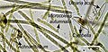
 Oceanides.v37i1-2.276-Fig2.jpg 475 × 598; 32 KB
-
 Oceanides.v37i1-2.276-Fig3.jpg 609 × 763; 34 KB
-
 Oocists de Cyclospora cayetanensis.png 1,561 × 1,872; 1.53 MB
-
 Osservazione al microscopio.jpg 960 × 1,280; 131 KB
-
 Paramecium undergoing Cyst Formation.jpg 4,160 × 3,120; 800 KB
-
 Parasite140076-fig1 Dirofilaria repens removed from a subcutaneous nodule - Photos.png 1,645 × 2,894; 5.31 MB
-
 Parasite170078-fig2 Cichlidogyrus philander (Monogenea, Ancyrocephalidae) (main image).png 2,107 × 1,035; 3.06 MB
-
 Parasite170078-fig2 Cichlidogyrus philander (Monogenea, Ancyrocephalidae).png 2,120 × 2,120; 6.46 MB
-
 Perlpolymer MB189.jpg 1,600 × 1,200; 344 KB
-
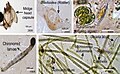
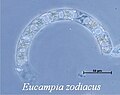 Photo de Eucampia zodiacus par microscopie optique.jpg 382 × 302; 29 KB
-
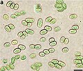
 Photo de Pseudo-nitzschia sp. par microscopie optique.jpg 385 × 302; 20 KB
-
 Physcomitrella patens LF (Protonema).jpg 2,099 × 1,763; 673 KB
-
 PhysRevLett.116.038102-Fig1.png 850 × 436; 303 KB
-
 PhysRevLett.116.038102-Fig1a.jpg 431 × 436; 40 KB
-
 Plants-11-00481-g002.png 2,010 × 2,931; 1.61 MB
-
 Plants-11-00481-g003.png 2,051 × 2,883; 1.42 MB
-
 Plphys v161 3 1334 f1-via-RG.jpg 850 × 780; 288 KB
-
 Plphys v161 3 1334 f1a-via-RG.jpg 277 × 237; 37 KB
-
 Plphys v161 3 1334 f1b-via-RG.jpg 278 × 238; 45 KB
-
 Plphys v161 3 1334 f1c-via-RG.jpg 277 × 239; 36 KB
-
 Plphys v161 3 1334 f1d-via-RG.jpg 277 × 236; 34 KB
-
 Plphys v161 3 1334 f1e-via-RG.jpg 276 × 236; 27 KB
-
 Plphys v161 3 1334 f1f-via-RG.jpg 277 × 232; 31 KB
-
 Pone.0118415.g001A.tif 411 × 1,490; 434 KB
-
 Protists (10.3897-subtbiol.42.78037) Figure 3D.jpg 435 × 393; 46 KB
-
 Protists (10.3897-subtbiol.42.78037) Figure 3E.jpg 270 × 558; 92 KB
-
 RFID Alien P5.jpg 6,571 × 6,518; 33.95 MB
-
 RFID NXP CUL1V2.jpg 3,174 × 3,091; 6.42 MB
-
 TORNAI-SpectrumOfMedicalImaging.jpg 720 × 504; 117 KB
-
 Toxins-16-00049-g001.png 3,123 × 2,351; 5.27 MB
-
 Toxins-16-00049-g003.png 3,014 × 2,270; 10.03 MB
-
 Vesicular Arbuscular Mycorrhizae 40X0031 03.jpg 2,592 × 1,944; 3.85 MB
-
 Комар под микроскопом.jpg 1,600 × 1,600; 149 KB


.jpg)


_(main_image).png)
.png)


.jpg)


_Figure_3D.jpg)
_Figure_3E.jpg)


