Category:Confocal microscopy
Jump to navigation
Jump to search
optical imaging technique for increasing optical resolution and contrast of a micrograph by means of using a spatial pinhole to block out-of-focus light in image formation | |||||
| Upload media | |||||
| Subclass of |
| ||||
|---|---|---|---|---|---|
| |||||
Subcategories
This category has the following 4 subcategories, out of 4 total.
Pages in category "Confocal microscopy"
This category contains only the following page.
Media in category "Confocal microscopy"
The following 17 files are in this category, out of 17 total.
-
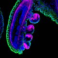
Colors of a tentacle.jpg 1,024 × 1,024; 171 KB
-
Confocal curve schematic.svg 464 × 329; 9 KB
-
Confocal of Hydrogel Podhorska.png 478 × 146; 95 KB
-
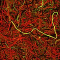
Immunofluorescence image of distributions of MBP and S100 in sciatic nerve of rat.png 1,024 × 1,024; 2.35 MB
-
Measuring-Principle-in-Confocal-Sensors.png 540 × 804; 100 KB
-
Microtubule binding domain fused with GFP-tag reveals microtubule network of HeLa cell line.jpg 12,788 × 12,788; 7.73 MB
-
Neuron in the microchannel of hydrogel implant.png 5,240 × 1,034; 9.97 MB
-
Paper Project image on Times Square New York.jpg 1,080 × 1,080; 1,021 KB
-
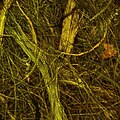
Sciatic nerve immunofluorescence image after implantation of silicone tube.png 1,024 × 1,024; 1,001 KB
-
Silk-handmade-paper-1280.jpg 1,280 × 1,280; 1.16 MB
-
Silk-handmade-paper-3d-1280.jpg 1,280 × 1,280; 1.09 MB
-
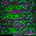
Spinal cord gray matter immunofluorescence staining, confocal imaging.png 1,600 × 1,600; 5.88 MB
-
-
Super-resolved Reflectance Confocal Microscopy 3D reconstruction of a Diatom shell - Zstack.webm 16 s, 840 × 420; 2.2 MB
-
Yucca-handmade-paper-1280.jpg 1,280 × 1,280; 1.3 MB
-
Yucca-handmade-paper-3d-1280.jpg 1,280 × 1,280; 1.53 MB
