File:Spider internal anatomy-en.svg
Jump to navigation
Jump to search

Size of this PNG preview of this SVG file: 800 × 351 pixels. Other resolutions: 320 × 140 pixels | 640 × 280 pixels | 1,024 × 449 pixels | 1,280 × 561 pixels | 2,560 × 1,122 pixels | 1,148 × 503 pixels.
Original file (SVG file, nominally 1,148 × 503 pixels, file size: 485 KB)
Captions
Captions
Add a one-line explanation of what this file represents
|

Summary
[edit]| Description |
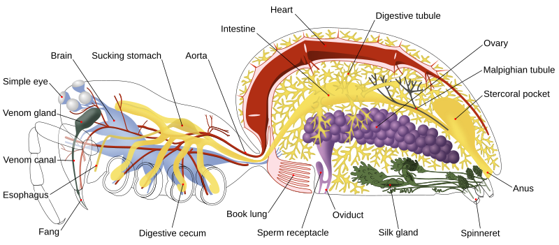
English: Diagram of the internal anatomy of a female two-lunged spider.
Français : Diagramme montrant l'anatomie interne d'une araignée femelle à deux poumons.
Deutsch: Diagramm der internen Anatomie einer weiblichen zweilungigen Spinne. Siehe File:Spider internal anatomy-de.svg. |
| Date | (UTC) |
| Source |
|
| Author |
Original: John Henry Comstock Vector: (Ryan Wilson) |
| Other versions |
Derivative works of this file: Spider anatomy blood.png
[]
|
| SVG development | This diagram was created with Inkscape. This diagram uses embedded text that can be easily translated using a text editor. |


This file is licensed under the Creative Commons Attribution 3.0 Unported license.
- You are free:
- to share – to copy, distribute and transmit the work
- to remix – to adapt the work
- Under the following conditions:
- attribution – You must give appropriate credit, provide a link to the license, and indicate if changes were made. You may do so in any reasonable manner, but not in any way that suggests the licensor endorses you or your use.
Original upload log
[edit]
This image comes from the following images:
- File:Spider_internal_anatomy.png licensed with PD-US
- 2009-06-27T01:33:45Z Kaldari 5000x2500 (10823835 Bytes) {{Information |Description={{en|1=Diagram of the internal anatomy of a two-lunged spider.}} |Source=Scanned from the 1920 edition of ''The Spider Book'', published by Doubleday, Page & Company (originally published in 1912)
Uploaded with derivativeFX
File history
Click on a date/time to view the file as it appeared at that time.
| Date/Time | Thumbnail | Dimensions | User | Comment | |
|---|---|---|---|---|---|
| current | 03:57, 9 January 2024 | | 1,148 × 503 (485 KB) | Kaldari (talk | contribs) | Fixing double line |
| 07:32, 19 April 2023 |  | 1,148 × 503 (443 KB) | Izaro.akk (talk | contribs) | File uploaded using svgtranslate tool (https://svgtranslate.toolforge.org/). Added translation for eu. | |
| 15:42, 2 March 2022 |  | 1,148 × 503 (438 KB) | Pho Sai (talk | contribs) | File uploaded using svgtranslate tool (https://svgtranslate.toolforge.org/). Added translation for my. | |
| 16:00, 26 October 2021 |  | 1,148 × 503 (438 KB) | Ninjastrikers (talk | contribs) | File uploaded using svgtranslate tool (https://svgtranslate.toolforge.org/). Added translation for my. | |
| 16:15, 30 November 2018 |  | 1,148 × 503 (432 KB) | Pbrks (talk | contribs) | optimized size | |
| 05:32, 22 May 2013 |  | 1,148 × 503 (636 KB) | Patrick Edwin Moran (talk | contribs) | Spiders produce venom in venom glands and deliver venom through venom canals, so two occurrences of the word "poison" were wrong and have now been connected. | |
| 05:27, 22 May 2013 |  | 1,148 × 503 (637 KB) | Patrick Edwin Moran (talk | contribs) | Spiders do not produce poison to use on prey. They produce venom from venom glands. So "poison gland" must be changed to "venom gland" to be correct. | |
| 07:33, 4 April 2011 |  | 1,148 × 503 (866 KB) | Kaldari (talk | contribs) | Removing coxal gland for 4 reasons: 1. Not present in Comstock's original diagram, of which this is supposed to be the vectorized version 2. It is not part of the circulatory system and thus should not be colored red 3. It is positioned incorrectly accord | |
| 11:50, 16 May 2010 |  | 1,148 × 503 (869 KB) | Justass (talk | contribs) | Reverted to version as of 18:51, 11 November 2009 | |
| 11:35, 16 May 2010 |  | 1,148 × 503 (902 KB) | Xvazquez (talk | contribs) | *Traslation to spanish *Traducción al español Category:Spider anatomy |
You cannot overwrite this file.
File usage on Commons
The following 40 pages use this file:
- SVG animal diagrams
- User:Pbrks/Images
- User talk:Pbrks/2008 July - 2011 May
- User talk:Pbrks/Archives/2009
- Commons:Featured picture candidates/File:Spider internal anatomy-en.svg
- Commons:Featured picture candidates/Log/November 2009
- Commons:Featured pictures/Non-photographic media/Computer-generated
- Commons:Featured pictures/chronological/2009-B
- Commons:Picture of the Year/2009/Galleries/2009-B
- Commons:Picture of the Year/2009/Galleries/All
- Commons:Picture of the Year/2009/Galleries/Diagrams
- Commons:Picture of the Year/2009/Galleries/Index/17
- Commons:Picture of the Year/2009/Galleries/Index/Diagrams
- Commons:Picture of the Year/2009/Galleries/Table/200911
- Commons:Picture of the Year/2009/R1/File:Spider internal anatomy-en.svg
- Commons:Picture of the Year/2009/Results/R1/ALL/Table
- Commons:Picture of the Year/2009/Results/R1/Diagrams
- Commons:Picture of the Year/2009/Results/R1/Diagrams/Table
- File:Spider anatomy.png
- File:Spider anatomy blood.png
- File:Spider internal anatomy-RO.png
- File:Spider internal anatomy-ar.svg
- File:Spider internal anatomy-cs.svg
- File:Spider internal anatomy-de.svg
- File:Spider internal anatomy-en.svg
- File:Spider internal anatomy-es.svg
- File:Spider internal anatomy-eu.svg
- File:Spider internal anatomy-fr.svg
- File:Spider internal anatomy-it.svg
- File:Spider internal anatomy-it Repro.png
- File:Spider internal anatomy-my.png
- File:Spider internal anatomy-my.svg
- File:Spider internal anatomy-numb.svg
- File:Spider internal anatomy-nv.svg
- File:Spider internal anatomy-sl.svg
- File:Spider internal anatomy-zh-Hant-TW.svg
- File:Spider internal anatomy.png
- File:Spider internal anatomy - altered description.jpg
- File:Spider internal anatomy PL.svg
- Template:Other versions/Spider internal anatomy
File usage on other wikis
The following other wikis use this file:
- Usage on af.wikipedia.org
- Usage on als.wikipedia.org
- Usage on ar.wikipedia.org
- Usage on az.wiktionary.org
- Usage on bg.wikipedia.org
- Usage on bs.wikipedia.org
- Usage on en.wikipedia.org
- Nervous system
- Lung
- Book lung
- Epigyne
- Wikipedia:WikiProject Spiders/Articles
- Spider anatomy
- Talk:Spider anatomy
- Portal:Arthropods/Selected picture
- Wikipedia:Featured pictures/Animals/Arachnids
- User talk:Pbrks/Archives/2009
- Wikipedia:Featured pictures thumbs/19
- Wikipedia:Featured picture candidates/August-2009
- Wikipedia:Graphics Lab/Image workshop/Archive/Aug 2009
- User talk:ZooFari/Archives/2009/August
- Wikipedia:Featured picture candidates/Spider internal anatomy
- Wikipedia:Wikipedia Signpost/2009-08-31/Features and admins
- User talk:Kaldari/Archive 7
- Wikipedia:Picture of the day/December 2010
- Spider
- Template:POTD/2010-12-13
- User:Venustas 12/December 13 2010/Main Page Picture
- User:Vietnamesepresident/Gallery
- User talk:Kaldari/Archive 9
- Pain in invertebrates
- User talk:Pbrks/Archives/2010
- Evolution of nervous systems
- Wikipedia:Wikipedia Signpost/2009-08-31/SPV
- User:Pbrks
- Wikipedia:Wikipedia Signpost/Single/2009-08-31
- Usage on eu.wikipedia.org
- Usage on fa.wikipedia.org
- Usage on he.wikipedia.org
- Usage on hi.wikipedia.org
- Usage on hu.wikipedia.org
- Usage on hy.wikipedia.org
- Usage on id.wikipedia.org
- Usage on it.wikipedia.org
- Usage on ja.wikipedia.org
- Usage on ka.wikipedia.org
View more global usage of this file.