Category:Transmission electron microscopy
Jump to navigation
Jump to search
analytic technique | |||||
| Upload media | |||||
| Subclass of | |||||
|---|---|---|---|---|---|
| Different from | |||||
| |||||
Subcategories
This category has the following 9 subcategories, out of 9 total.
Media in category "Transmission electron microscopy"
The following 86 files are in this category, out of 86 total.
-
1-s2.0-S215075112400474X-mbio.01623-24.f002B+.gif 360 × 178; 35 KB
-
2021.03.30.437421v4.S3.jpg 868 × 866; 102 KB
-
2021.03.30.437421v4.S5.jpg 1,869 × 919; 494 KB
-
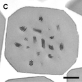
2021.03.30.437421v4.S6ABC.jpg 1,357 × 898; 133 KB
-
2021.03.30.437421v4.S7.jpg 886 × 885; 90 KB
-
41467 2023 39758 Fig1.webp 1,494 × 1,212; 472 KB
-
41467 2023 39758 Fig1f.jpg 453 × 454; 125 KB
-
Autonomous-beating-rate-adaptation-in-human-stem-cell-derived-cardiomyocytes-ncomms10312-s2.ogv 8.7 s, 640 × 480; 600 KB
-
Autonomous-beating-rate-adaptation-in-human-stem-cell-derived-cardiomyocytes-ncomms10312-s3.ogv 13 s, 640 × 480; 2.59 MB
-
Autonomous-beating-rate-adaptation-in-human-stem-cell-derived-cardiomyocytes-ncomms10312-s4.ogv 14 s, 640 × 480; 1.3 MB
-
-
BIOC - Laboratoire de Biochimie (16867498367).jpg 5,760 × 3,840; 17.14 MB
-
Colloidal Gold Labeling (8530568499).jpg 450 × 285; 32 KB
-
-
-
-
-
-
-
-
-
-
Direct-observation-of-catalytic-oxidation-of-particulate-matter-using-in-situ-TEM-srep10161-s3.ogv 1 min 11 s, 640 × 480; 1.08 MB
-
-
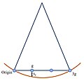
Ewald Sphere Weak-beam Dark-field 3g Conditions.jpg 615 × 589; 27 KB
-
Examples of Embedded and Sectioned Material (8531814250).jpg 856 × 226; 116 KB
-
FEI Vitrobot (8510271991).jpg 700 × 449; 187 KB
-
FEI-Tecnai BT Spirit TEM (8531837620).jpg 450 × 597; 61 KB
-
Fib sample preparation tem.jpg 2,400 × 800; 827 KB
-
Fibres-and-cellular-structures-preserved-in-75-million–year-old-dinosaur-specimens-ncomms8352-s2.ogv 33 s, 983 × 686; 12.02 MB
-
Fibres-and-cellular-structures-preserved-in-75-million–year-old-dinosaur-specimens-ncomms8352-s3.ogv 24 s, 1,100 × 565; 15.39 MB
-
Freeze Fracture Replicas (8512437771).jpg 540 × 378; 60 KB
-
Gaas inas quantum dot.jpg 1,400 × 484; 550 KB
-
-
-
-
-
-
-
Light and Electron Micrographs (8516088750).jpg 260 × 132; 15 KB
-
Maturation-of-the-HIV-1-core-by-a-non-diffusional-phase-transition-ncomms6854-s2.ogv 6.3 s, 1,280 × 720; 11.83 MB
-
Maturation-of-the-HIV-1-core-by-a-non-diffusional-phase-transition-ncomms6854-s3.ogv 16 s, 939 × 858; 8.31 MB
-
Mbio.00574-21-f001.jpg 1,800 × 1,376; 607 KB
-
Mbio.00574-21-f001a.jpg 486 × 365; 76 KB
-
Mechanism-of-cellular-uptake-of-genotoxic-silica-nanoparticles-1743-8977-9-29-S7.ogv 9.3 s, 640 × 480; 6.76 MB
-
Mechanism-of-cellular-uptake-of-genotoxic-silica-nanoparticles-1743-8977-9-29-S8.ogv 9.3 s, 640 × 480; 4.83 MB
-
-
ODD.Baculo.Fig4.v1.png 1,778 × 1,770; 816 KB
-
ODD.Baculo.Fig4C.v1.png 867 × 870; 276 KB
-
ODD.Baculo.Fig4D.v1.png 866 × 860; 396 KB
-
Parasite (journal) 2013, 20, 28 Miquel - Pleurogenidae.pdf 1,233 × 1,747, 15 pages; 12.19 MB
-
Parasite140105-fig3 Toxoplasmosis in a bar-shouldered dove - TEM of 2 tachyzoites.tif 1,378 × 1,683; 2.11 MB
-
Parasite150063-fig3 Spermatozoon of Atractotrema sigani (Digenea) TEM.tif 2,343 × 2,198, 2 pages; 7.59 MB
-
PE Bruchfläche.jpg 857 × 845; 342 KB
-
Perovskite oxide thin film.jpg 1,400 × 1,272; 646 KB
-
Schematic view of imaging and diffraction modes in TEM..tif 4,079 × 3,654; 42.64 MB
-
Secretory-glands-in-cercaria-of-the-neuropathogenic-schistosome-Trichobilharzia-regenti--1756-3305-4-162-S1.ogv 8.3 s, 1,120 × 832; 3.18 MB
-
Secretory-glands-in-cercaria-of-the-neuropathogenic-schistosome-Trichobilharzia-regenti--1756-3305-4-162-S2.ogv 8.3 s, 1,120 × 832; 7.38 MB
-
-
TEM image of Pt polycrystalline film and correspondent diffraction pattern.tif 1,914 × 1,311; 7.18 MB
-
TEM-micrographs-of-2wt.jpg 660 × 279; 112 KB
-
Tensile-Forces-Originating-from-Cancer-Spheroids-Facilitate-Tumor-Invasion-pone.0156442.s015.ogv 2.0 s, 1,024 × 1,024; 2.56 MB
-
Tensile-Forces-Originating-from-Cancer-Spheroids-Facilitate-Tumor-Invasion-pone.0156442.s016.ogv 15 s, 872 × 872; 1.89 MB
-
The neuroblast of Drosophila melanogaster.tif 2,048 × 2,048; 4 MB
-
-
-
The-Notch-and-Wnt-pathways-regulate-stemness-and-differentiation-in-human-fallopian-tube-organoids-ncomms9989-s4.ogv 1 min 12 s, 959 × 1,076; 5.11 MB
-
-
-
Trans-mitochondrial-coordination-of-cristae-at-regulated-membrane-junctions-ncomms7259-s2.ogv 16 s, 650 × 728; 22.89 MB
-
Transmission Electron Microscope operating principle.ogv 2 min 32 s, 1,920 × 1,080; 11.28 MB
-
Transmission electron microscopy, Laguna Negra.jpg 851 × 498; 319 KB
-
Type-II-spiral-ganglion-afferent-neurons-drive-medial-olivocochlear-reflex-suppression-of-the-ncomms8115-s2.ogv 9.7 s, 1,280 × 720; 7.43 MB
-
Type-II-spiral-ganglion-afferent-neurons-drive-medial-olivocochlear-reflex-suppression-of-the-ncomms8115-s3.ogv 9.7 s, 1,280 × 720; 5.33 MB
-
-
-
-
-
-
Weak-Beam Dark-Field Example Image 1.5kx Fringes.jpg 512 × 512; 54 KB
-
Weak-Beam Dark-Field Example Image 1.5kx.jpg 512 × 512; 67 KB
-
Weak-Beam Dark-Field Example Image 2kx Fringes.jpg 512 × 512; 59 KB
-
Weak-beam dark-field TEM 3g diffraction conditions.jpg 1,132 × 553; 38 KB
-
Weak-beam dark-field TEM image of dislocations.jpg 1,280 × 720; 112 KB
-
Ежик в тумане.jpg 1,000 × 777; 136 KB


