Category:Transmission electron microscopic images
Jump to navigation
Jump to search
Subcategories
This category has the following 8 subcategories, out of 8 total.
C
E
Media in category "Transmission electron microscopic images"
The following 139 files are in this category, out of 139 total.
-
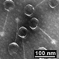
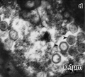
12985 2018 930 Fig4 HTML (d) Cedratvirus.png 130 × 120; 14 KB
-
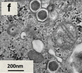
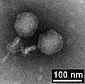
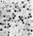
12985 2018 930 Fig4 HTML (f) Marseillevirus.png 136 × 121; 17 KB
-
41396 2019 362 Fig1 HTML.webp 1,911 × 734; 99 KB
-
41396 2019 362 Fig1B ICBM1.jpg 344 × 334; 46 KB
-
41396 2019 362 Fig1B ICBM2.jpg 342 × 333; 62 KB
-
41598 2013 Article BFsrep03337 Fig2 HTML (F).png 1,011 × 758; 1.08 MB
-
41598 2013 Article BFsrep03337 Fig2 HTML.png 2,035 × 2,287; 5.17 MB
-
6816k240zones.png 256 × 256; 81 KB
-
A red blood cell in a capillary, pancreatic tissue - TEM.jpg 1,560 × 1,254; 579 KB
-
Alpha-mannosidosis electron micrograph.JPEG 839 × 439; 76 KB
-
AML-M5B, promonocyte, TEM.jpg 685 × 512; 309 KB
-
Ancient DNA.png 606 × 594; 157 KB
-
Asp Phyllokladius.jpg 1,360 × 1,032; 180 KB
-
Braarudosphaera bigelowii TEM.png 768 × 856; 438 KB
-
CagedSpider.png 306 × 244; 70 KB
-
Chloride cell.jpg 574 × 480; 276 KB
-
Cilia lobostoma2.jpg 1,300 × 981; 461 KB
-
Cilia.jpg 640 × 455; 75 KB
-
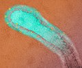
Colorized transmission electron micrograph of monkeypox virus particles (green).jpg 2,915 × 2,252; 2.81 MB
-
Desmosome - epithelial cell from mammalian lung tissue - TEM.jpg 640 × 480; 148 KB
-
DifraccionElectronesMET.jpg 354 × 354; 63 KB
-
E. coli fimbriae.png 649 × 832; 326 KB
-
Eaay9634.F1B.large.jpg 374 × 380; 94 KB
-
ER-containing autophagosome.png 1,417 × 1,770; 2.06 MB
-
F21-01-9780123846846 mimivirus particle stargate.png 1,642 × 1,400; 1.2 MB
-
F21-02-9780123846846 mimivirus particle internals.png 1,642 × 1,552; 1.24 MB
-
F21-04-9780123846846 mimivirus virion factory.png 1,867 × 2,241; 1.92 MB
-
Fig3-TEM-micrographs-of-samples.gif 711 × 546; 334 KB
-
Fivefoldtwin.png 664 × 374; 235 KB
-
Fmicb-05-00506-g001.jpg 943 × 952; 257 KB
-
Fmicb-05-00506-g001B.jpg 442 × 455; 62 KB
-
Fmicb-05-00506-g001D.jpg 433 × 449; 64 KB
-
Fmicb-07-01740-g002.jpg 964 × 970; 678 KB
-
Fmicb-07-01740-g002a extr.jpg 335 × 194; 54 KB
-
Fmicb-07-01740-g002a.jpg 473 × 316; 89 KB
-
Fmicb-07-01740-g002e.jpg 474 × 318; 71 KB
-
Fmicb-07-01740-g003.jpg 964 × 335; 286 KB
-
Fmicb-08-02659-g001.jpg 1,928 × 978; 172 KB
-
Fmicb-08-02659-g001B-15497.jpg 451 × 449; 23 KB
-
Fmicb-08-02659-g001B-15519.jpg 442 × 443; 20 KB
-
Fmicb-08-02659-g001B-15599.jpg 454 × 447; 26 KB
-
Fmicb-08-02659-g001B-15708.jpg 450 × 444; 27 KB
-
Fpls-06-01009-g001.jpg 632 × 652; 130 KB
-
Glittering Lead Nanoparticles.tif 1,024 × 768; 772 KB
-
His1 MDS 2011.jpg 287 × 248; 61 KB
-
Hohllatex1.jpg 2,511 × 1,797; 418 KB
-
Hohllatex2.jpg 2,364 × 926; 227 KB
-
Hohllatex3.jpg 1,024 × 768; 89 KB
-
Hrp Labeling (8531659348).jpg 256 × 159; 6 KB
-
HTREM atoms.jpg 1,650 × 1,200; 1.31 MB
-
Human leukocyte, showing golgi - TEM.jpg 640 × 480; 97 KB
-
Human Respiratory Syncytial Virus (RSV) (52453014952).jpg 1,892 × 1,576; 1.02 MB
-
Icogdfam.gif 620 × 620; 209 KB
-
Ijms-21-02096-g001.webp 2,211 × 1,102; 761 KB
-
Ijms-21-02096-g001a.jpg 1,071 × 1,071; 357 KB
-
Ijms-21-02096-g001b.jpg 1,071 × 1,071; 346 KB
-
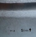
Kikuchi lines Si.png 806 × 794; 230 KB
-
Latex tem1.jpg 1,600 × 1,000; 164 KB
-
Latex tem2.jpg 2,424 × 1,719; 474 KB
-
LegionellaPneumophila.jpg 617 × 499; 78 KB
-
MBio-2020-Subramaniam-e02938-19.F4 (D-I).large.TMEs.jpg 1,280 × 810; 306 KB
-
MBio-2020-Subramaniam-e02938-19.F5.large.TMEjpg.jpg 1,280 × 790; 307 KB
-
MCV VLP EM PTA staining.jpg 1,623 × 1,797; 596 KB
-
Methylococcus capsulatus.png 600 × 591; 184 KB
-
Microorganisms-08-00400-g002.png 2,700 × 1,364; 1.01 MB
-
Microorganisms-08-00400-g002B.png 772 × 597; 306 KB
-
NiSi2 Si interface crop.png 800 × 445; 233 KB
-
Norwalk.jpg 229 × 238; 12 KB
-
NuclearTrackPaper02chromiteA.jpg 1,188 × 944; 402 KB
-
ODD.Baculo.Fig1.v3.png 3,801 × 5,187; 6.2 MB
-
OPSR.Sarthro.Fig1.v1.png-640x480.png 1,280 × 498; 582 KB
-
OPSR.Sarthro.Fig1l.v1.png-640x480.png 282 × 189; 58 KB
-
OPSR.Sarthro.Fig1r.v1.png-640x480.png 317 × 249; 94 KB
-
Pancreatic cells - TEM.jpg 640 × 480; 139 KB
-
Parasite140019-fig1 Nosema podocotyloidis - Hyperparasitic Microsporidia.tif 2,022 × 3,031; 4 MB
-
Parasite140019-fig2 Nosema podocotyloidis - Hyperparasitic Microsporidia.tif 2,453 × 2,585; 3.74 MB
-
Parasite140019-fig3 Nosema podocotyloidis - Hyperparasitic Microsporidia.tif 2,453 × 2,204; 3.68 MB
-
Parasite140019-fig4 Nosema podocotyloidis - Hyperparasitic Microsporidia.tif 2,453 × 2,531; 3.14 MB
-
Parasite140083-fig6 Figs 37-44 Cathayacanthus spinitruncatus.tif 2,067 × 2,499; 3.42 MB
-
Parasite170127-fig4 ovipositor Pimplinae TEM.png 2,236 × 2,521; 9.65 MB
-
Parasite170127-fig5 ovipositor Pimplinae TEM.png 2,331 × 2,624; 9.23 MB
-
Parasite170127-fig6 ovipositor Pimplinae.png 2,814 × 2,109; 9.06 MB
-
Parasite170144-fig17 Phylogenetic trees of Polyopisthocotylea.png 8,188 × 3,783; 827 KB
-
Pone.0061912.g003 (B) Sputnik3 TEM(-).tif 404 × 303; 186 KB
-
Pone.0061912.g003 (C) Sputnik3 InCulture.tif 402 × 322; 181 KB
-
PRV Virion Feature.png 608 × 524; 256 KB
-
PRV Virion.png 2,242 × 591; 1.57 MB
-
Pt on ssDNA on NTs.jpg 1,024 × 1,024; 465 KB
-
Respiratory syncytial virus 01.jpg 1,804 × 1,196; 1.23 MB
-
Root-nodule01.jpg 2,240 × 1,654; 2.3 MB
-
Rotavirus infected gut.jpg 666 × 732; 86 KB
-
Sbs block copolymer.jpg 1,018 × 717; 257 KB
-
Science & Nature.jpg 2,048 × 2,048; 2.48 MB
-
See the Light.jpg 1,823 × 1,823; 3.25 MB
-
Septatejunction.jpg 369 × 393; 60 KB
-
Simulation GaN.png 455 × 684; 55 KB
-
TEM - Epixenosomes.jpg 556 × 526; 110 KB
-
TEM image of ZIF-8.tif 2,048 × 2,048; 12 MB
-
TEM images of silica-organic matrix-based suspension.jpg 731 × 397; 269 KB
-
TEM nanotubos.jpg 432 × 217; 20 KB
-
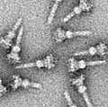
TEM of isolated T3SS needle complexes.jpg 271 × 269; 53 KB
-
TEM of silicon nanowire (5887830328).jpg 1,800 × 1,677; 1,023 KB
-
TEM Orientation Mapping.png 1,117 × 703; 889 KB
-
TEM phil CDC 11635 lores.jpg 700 × 748; 137 KB
-
TEM-image-for-CSNC2-4wt.jpg 600 × 600; 161 KB
-
TEM-image-of-GO.jpg 600 × 601; 193 KB
-
TEM-image-of-nanocrystalline-Ni95Ti5-catalyst.jpg 600 × 483; 31 KB
-
TEM-images-of-mesoporous-Ta-sub3sub-W-sub7sub-oxide-stearic-acid.jpg 660 × 633; 333 KB
-
TEM-photograph-of-CeO2-nanopowder.jpg 600 × 494; 51 KB
-
TEM-Versetzungen.png 611 × 540; 66 KB
-
Tight junction blowup.jpg 882 × 751; 389 KB
-
TiMnImage2y.png 512 × 512; 460 KB
-
Titanium dioxide nanoparticles.png 434 × 265; 134 KB
-
Two desmosomes - TEM.jpg 640 × 480; 111 KB
-
Ultrastructure of choroid epithelium.jpg 1,200 × 1,000; 414 KB
-
Ultrastructure of tracheal hemidesmosomes in mice.JPEG 994 × 1,200; 350 KB
-
Vibrio cholerae.jpg 1,762 × 1,437; 1.61 MB
-
Viruses-09-00046-g002a Pithovirus.jpg 2,400 × 1,465; 676 KB
-
Viruses-11-00312-g003.png 1,454 × 1,918; 1.35 MB
-
Viruses-11-00312-g003B.png 680 × 739; 330 KB
-
Viruses-11-00312-g003C.png 1,395 × 1,087; 1.08 MB
-
Viruses-11-00440-g002.webp 2,579 × 1,423; 1.2 MB
-
Viruses-11-00440-g002A.png 1,253 × 1,345; 1.19 MB
-
Viruses-11-00440-g002B.png 1,242 × 1,347; 1.3 MB
-
Zemlin-Tableau after correction of axial coma.jpg 680 × 680; 200 KB
-
Zemlin-Tableau before correction of axial coma.jpg 680 × 680; 192 KB

