Category:Human sacrum
Appearance
| Human Vertebral column | |||||||||||
|
 Human anatomy Sacrum
| ||
|---|---|---|
triangular-shaped bone at the bottom of the spine | |||||
| Upload media | |||||
| Instance of |
| ||||
|---|---|---|---|---|---|
| Subclass of |
| ||||
| Part of |
| ||||
| Connects with |
| ||||
| Has part(s) |
| ||||
| Different from | |||||
| |||||
Subcategories
This category has the following 7 subcategories, out of 7 total.
3
- 3D data of human sacrum (2 F)
A
O
P
- Photographs of human sacrum (36 F)
V
- Videos of human sacrum (2 F)
Media in category "Human sacrum"
The following 123 files are in this category, out of 123 total.
-
720 Sacrum and Coccyx.jpg 1,186 × 530; 304 KB
-
Anterior surface of sacrum.jpg 960 × 720; 106 KB
-
Base of the spine and hip bones.jpg 1,390 × 1,536; 389 KB
-
BodyParts3D FJ6484 Sacrum.stl 5,120 × 2,880; 481 KB
-
Braus 1921 50.png 916 × 2,520; 6.62 MB
-
Braus 1921 79.png 496 × 600; 874 KB
-
Bulletin (1916) (19802814973).jpg 1,712 × 2,460; 1.05 MB
-
Congenital fistula over the sacrum Wellcome L0062487.jpg 5,372 × 3,804; 3.15 MB
-
Congenital sacral tumour in a man Wellcome L0066982.jpg 3,852 × 5,204; 1.3 MB
-
Congenital sacral tumour in a man Wellcome L0066983.jpg 3,955 × 5,306; 1.36 MB
-
Cunningham’s Text-book of Anatomy (1914) - Fig 120.png 808 × 561; 468 KB
-
Cunningham’s Text-book of Anatomy (1914) - Fig 395.png 1,680 × 1,614; 2.34 MB
-
Decubitus 01.JPG 1,024 × 768; 376 KB
-
Dem bones (3387445).jpg 1,945 × 1,536; 1.24 MB
-
Dixon's Manual of human osteology (1912) - Fig 013.png 1,287 × 1,584; 1.32 MB
-
Dixon's Manual of human osteology (1912) - Fig 014.png 1,278 × 1,605; 787 KB
-
Dixon's Manual of human osteology (1912) - Fig 015.png 1,312 × 1,388; 1.25 MB
-
Dixon's Manual of human osteology (1912) - Fig 016.png 1,320 × 717; 636 KB
-
Enormously large congenital sacral tumour.jpg 1,613 × 1,705; 588 KB
-
Gerrish's Text-book of Anatomy (1902) - Fig. 141.png 1,134 × 1,073; 859 KB
-
Gerrish's Text-book of Anatomy (1902) - Fig. 142.png 1,288 × 1,148; 939 KB
-
Gerrish's Text-book of Anatomy (1902) - Fig. 143.png 1,416 × 1,887; 1.58 MB
-
Gray 111 - Vertebral column-coloured Ro (3) mod.JPG 259 × 147; 9 KB
-
Gray 111 - Vertebral column-coloured Ro (3).JPG 259 × 147; 8 KB
-
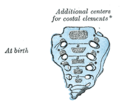
Gray107.png 277 × 236; 9 KB
-
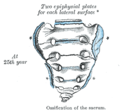
Gray108.png 283 × 226; 12 KB
-
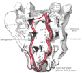
Gray109.png 338 × 312; 12 KB
-
Gray110.png 405 × 212; 11 KB
-
Gray1213.png 250 × 500; 18 KB
-
Gray322.png 518 × 307; 29 KB
-
Gray323.png 486 × 308; 26 KB
-
Gray324.png 400 × 205; 17 KB
-
Gray803.png 576 × 500; 100 KB
-
Gray95-ar.png 525 × 550; 157 KB
-
Gray95.png 525 × 550; 70 KB
-
Gray96-ar.png 567 × 500; 105 KB
-
Gray96.png 567 × 500; 45 KB
-
Gray97-ar.png 381 × 600; 89 KB
-
Gray97.png 381 × 600; 40 KB
-
Gray98 zh.png 500 × 333; 163 KB
-
Gray98-ar.png 500 × 333; 92 KB
-
Gray98.png 500 × 333; 42 KB
-
Gray99.png 317 × 550; 24 KB
-
HK TST Science Museum Bones exhibit 23 人類 skeletons.JPG 2,448 × 3,264; 1.61 MB
-
Holden's human osteology (1899) - Plt28 Fig01.png 1,035 × 1,005; 558 KB
-
Holden's human osteology (1899) - Plt28 Fig02.png 1,650 × 1,002; 992 KB
-
Holden's human osteology (1899) - Plt29.png 1,761 × 1,761; 1.42 MB
-
Holden's human osteology (1899) - Plt43.png 1,038 × 3,077; 1.35 MB
-
Image-Ass 2 - Sacrum.png 405 × 541; 119 KB
-
Kryžkaulio dubeninis paviršius.png 624 × 384; 118 KB
-
Kryžkaulio nugarinis paviršius.png 624 × 359; 128 KB
-
Merkel's Human Anatomy (1913) - Vol 3 - Fig 118.png 1,664 × 2,208; 675 KB
-
Mines de Gavà 023.JPG 3,648 × 2,736; 3.4 MB
-
Morris' human anatomy (1898) - Fig 017.png 1,491 × 1,335; 1.27 MB
-
Morris' human anatomy (1898) - Fig 018.png 1,620 × 1,104; 1.3 MB
-
Morris' human anatomy (1933) - Fig 106.png 1,947 × 1,650; 2.04 MB
-
Morris' human anatomy (1933) - Fig 107.png 2,040 × 1,356; 2.34 MB
-
Pelvis - os sacrum (anterior).jpg 2,460 × 3,048; 3.43 MB
-
Pelvis - os sacrum (caudal view).jpg 3,844 × 2,492; 4.26 MB
-
Pelvis - os sacrum (lateral view).jpg 3,692 × 2,320; 3.71 MB
-
Pelvis - os sacrum (posterior) 2.jpg 3,720 × 2,496; 3.71 MB
-
Pelvis - os sacrum (posterior).jpg 2,748 × 3,192; 3.18 MB
-
Piersol's human anatomy (1919) - Fig. 152.png 1,450 × 1,458; 2.36 MB
-
Piersol's human anatomy (1919) - Fig. 153.png 1,520 × 1,552; 1.78 MB
-
Piersol's human anatomy (1919) - Fig. 154.png 1,022 × 1,370; 1,010 KB
-
Posterior surface of sacrum.jpg 960 × 720; 123 KB
-
Sacral region of spinal cord.gif 193 × 357; 5 KB
-
Sacrum (Filled Holes).stl 5,120 × 2,880; 46.38 MB
-
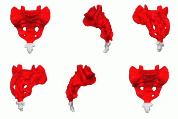
Sacrum - animation00.gif 360 × 360; 2.41 MB
-
Sacrum - animation01.gif 360 × 360; 2.56 MB
-
Sacrum - animation02.gif 360 × 360; 4.1 MB
-
Sacrum - animation03.gif 360 × 360; 4.76 MB
-
Sacrum - animation04.gif 360 × 360; 2.37 MB
-
Sacrum - animation05.gif 360 × 360; 1.65 MB
-
Sacrum - animation06.gif 360 × 360; 1.87 MB
-
Sacrum - anterior view00.png 1,125 × 1,125; 426 KB
-
Sacrum - anterior view01.png 1,125 × 1,125; 215 KB
-
Sacrum - anterior view02.png 1,125 × 1,125; 421 KB
-
Sacrum - anterior view03.png 1,125 × 1,125; 459 KB
-
Sacrum - anterior view04.png 1,125 × 1,125; 341 KB
-
Sacrum - anterior view05.png 1,125 × 1,125; 298 KB
-
Sacrum - anterior view06.png 1,125 × 1,125; 330 KB
-
Sacrum - inferior view01.png 1,125 × 1,125; 276 KB
-
Sacrum - lateral view00.png 1,125 × 1,125; 247 KB
-
Sacrum - lateral view01.png 1,125 × 1,125; 129 KB
-
Sacrum - lateral view02.png 1,125 × 1,125; 208 KB
-
Sacrum - lateral view03.png 1,125 × 1,125; 249 KB
-
Sacrum - lateral view04.png 1,125 × 1,125; 194 KB
-
Sacrum - lateral view05.png 1,125 × 1,125; 185 KB
-
Sacrum - posterior view00.png 1,125 × 1,125; 438 KB
-
Sacrum - posterior view01.png 1,125 × 1,125; 220 KB
-
Sacrum - posterior view02.png 1,125 × 1,125; 457 KB
-
Sacrum - posterior view03.png 1,125 × 1,125; 499 KB
-
Sacrum - posterior view04.png 1,125 × 1,125; 345 KB
-
Sacrum - posterior view05.png 1,125 × 1,125; 340 KB
-
Sacrum - posterior view06.png 1,125 × 1,125; 348 KB
-
Sacrum - superior view01.png 1,125 × 1,125; 282 KB
-
Sacrum .tif 976 × 505; 193 KB
-
Sacrum 1300283.JPG 2,304 × 3,072; 2.34 MB
-
Sacrum Anatomy by Jason Christian.webm 1 min 12 s, 1,280 × 720; 11.14 MB
-
Sacrum and Coccyx Overview.webm 1 min 23 s, 1,077 × 606; 9.64 MB
-
Sacrum.png 680 × 642; 178 KB
-
Sacrum1.png 667 × 663; 276 KB
-
Slide1gt.JPG 960 × 720; 129 KB
-
Slide2CORO-ar.jpg 960 × 720; 119 KB
-
Slide2CORO.JPG 960 × 720; 59 KB
-
Slide4CORO-ar.jpg 960 × 720; 216 KB
-
Slide4CORO.JPG 960 × 720; 113 KB
-
Slide8BLA.JPG 960 × 720; 97 KB
-
Sobo 1909 13-ar.png 1,764 × 1,636; 1.71 MB
-
Sobo 1909 13.png 1,764 × 1,636; 8.27 MB
-
Sobo 1909 14.png 1,472 × 1,560; 6.58 MB
-
Sobo 1909 15.png 1,668 × 884; 5.63 MB
-
Sobo 1909 16.png 1,288 × 748; 2.76 MB
-
Sobo 1909 17-ar.png 1,100 × 1,580; 1,008 KB
-
Sobo 1909 17.png 1,100 × 1,580; 4.98 MB
-
Sobo 1909 18.png 1,168 × 1,692; 5.66 MB
-
Sobo 1909 210.png 2,188 × 1,724; 10.81 MB
-
Sobo 1909 215.png 2,456 × 1,696; 11.94 MB
-
Testut's Treatise on Human Anatomy (1911) - Vol 1 - Fig 093.png 1,080 × 1,326; 680 KB
-
Testut's Treatise on Human Anatomy (1911) - Vol 1 - Fig 094.png 1,047 × 1,206; 1 MB
-
The science and art of midwifery (1897) (14760387811).jpg 2,036 × 3,044; 441 KB


