
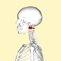
Category:Human atlas
| Human Vertebral column | |||||||||||
|
Subcategories
This category has the following 7 subcategories, out of 7 total.
Media in category "Human atlas"
The following 72 files are in this category, out of 72 total.
-
 Arcusposterioratlantis.png 500 × 235; 18 KB
-
 Atlante (C1) inf.JPG 2,432 × 1,824; 469 KB
-
 Atlante (C1).JPG 2,432 × 1,824; 482 KB
-
 Atlas (C1) animation small.gif 320 × 320; 859 KB
-
 Atlas (C1) back.png 900 × 900; 187 KB
-
 Atlas (C1) from top animation small.gif 320 × 320; 1.53 MB
-
 Atlas (C1) lateral.png 900 × 900; 166 KB
-
 Atlas (son).tif 991 × 496; 140 KB
-
 Atlas .tif 1,000 × 497; 140 KB
-
 Atlas Bone and Spine.gif 1,280 × 720; 19.79 MB
-
 Atlas Bone.gif 1,280 × 720; 5.43 MB
-
 Atlas bone.jpg 1,280 × 720; 62 KB
-
 Atlas Gr a.png 500 × 400; 31 KB
-
 Atlas The First Cervical Vertebra.gif 800 × 800; 7.15 MB
-
 Atlas vertebra inferior view.jpg 1,224 × 1,014; 92 KB
-
 Atlas vertebra superior sight.jpg 1,224 × 1,014; 101 KB
-
 Atlas vertebrae.jpg 960 × 720; 69 KB
-
 Atlasbogenspalt.jpg 1,142 × 1,031; 110 KB
-
 AtlasBone Image.jpg 1,280 × 720; 108 KB
-
 BodyParts3D FJ6295 Atlas.stl 5,120 × 2,880; 100 KB
-
 Braus 1921 70.png 1,344 × 1,227; 4.73 MB
-
 Braus 1921 71.png 732 × 411; 883 KB
-
 Braus 1921 72.png 1,179 × 678; 2.29 MB
-
 C1 back.png 900 × 900; 360 KB
-
 C1 lateral.png 4,500 × 4,500; 3.61 MB
-
 C1-C2 AP.JPG 2,303 × 1,800; 382 KB
-
 C1-C2 Lat.JPG 2,470 × 1,714; 302 KB
-
 Cambridge Natural History Mammalia Fig 007.png 416 × 261; 12 KB
-
 Cervical vertebra 1 animation bottom.gif 600 × 600; 1.26 MB
-
 Cervical vertebra 1 animation lateral.gif 600 × 600; 901 KB
-
 Cervical vertebra 1 animation top.gif 600 × 600; 1.31 MB
-
 Cervical vertebra 1 close-up back.png 900 × 900; 70 KB
-
 Cervical vertebra 1 close-up bottom.png 900 × 900; 101 KB
-
 Cervical vertebra 1 close-up frontal.png 900 × 900; 57 KB
-
 Cervical vertebra 1 close-up lateral.png 900 × 900; 47 KB
-
 Cervical vertebra 1 close-up top.png 900 × 900; 102 KB
-
 Condylar canal.jpg 708 × 518; 568 KB
-
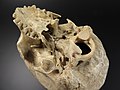 Cranium - atlas junction.jpg 4,608 × 3,456; 3.32 MB
-
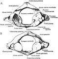
 Cunningham’s Text-book of Anatomy (1914) - Fig 119.png 1,740 × 782; 850 KB
-
De-Atlas.ogg 1.2 s; 12 KB
-
 Dixon's Manual of human osteology (1912) - Fig 006.png 1,194 × 1,218; 356 KB
-
 Gerrish's Text-book of Anatomy (1902) - Fig. 134.png 1,148 × 599; 399 KB
-
 Gray104.png 394 × 150; 5 KB
-
 Gray305.png 575 × 504; 50 KB
-
 Gray306.png 450 × 250; 18 KB
-
 Gray307.png 600 × 416; 53 KB
-
 Gray308.png 500 × 485; 51 KB
-
 Gray387.png 443 × 600; 54 KB
-
 Gray800.png 600 × 382; 52 KB
-
 Gray86 zh.png 500 × 235; 45 KB
-
 Gray86-ar.png 910 × 428; 261 KB
-
 Gray86.heb.PNG 500 × 235; 14 KB
-
 Gray86.png 500 × 235; 17 KB
-
 Human Atlas.stl 5,120 × 2,880; 4.35 MB
-
 HWS seitlich Annotation.jpg 754 × 1,100; 80 KB
-
 Inkompletter Ponticulus HWK 1 - Roe seitlich - 001 - Annotation.png 714 × 977; 547 KB
-
 Inkompletter Ponticulus HWK 1 - Roe seitlich - 001.png 714 × 977; 450 KB
-
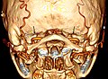
 Jeffersonfraktur - 84jm- CT axial - 001.jpg 1,826 × 1,128; 181 KB
-
 Posterior arch defect.png 718 × 454; 379 KB
-
 Sobo 1909 186.png 1,192 × 992; 3.39 MB
-
 Sobo 1909 187.png 1,256 × 1,116; 4.02 MB
-
 Sobo 1909 188.png 1,344 × 1,048; 4.04 MB
-
 Sobo 1909 243.png 2,100 × 1,820; 10.95 MB
-
 Sobo 1909 7.png 1,716 × 1,004; 4.94 MB
-

-
 Transverse foramen - atlas1.png 650 × 455; 283 KB
-
 Transverse foramen - atlas2.png 619 × 417; 236 KB
-
 Transverse foramen - atlas3.png 650 × 433; 252 KB
-
 Transverse foramen - atlas4.png 650 × 488; 406 KB
-
 Vertebra - atlas junction.jpg 4,608 × 3,456; 3.1 MB
-
 Vertebra - atlas.jpg 3,080 × 2,404; 587 KB
-
 Virchow - Menschen- und Affenschädel - 10 b1.png 448 × 381; 43 KB

_inf.JPG)
.JPG)
_animation_small.gif)
_back.png)
_from_top_animation_small.gif)
_lateral.png)


_-_Fig_119.png)
_-_Fig_006.png)
_-_Fig._134.png)


_(14776278441).jpg)


