Category:Bones of the human pelvis
Jump to navigation
Jump to search
 Human anatomy Bones of the pelvis
| ||
|---|---|---|
Subcategories
This category has the following 8 subcategories, out of 8 total.
A
C
F
H
P
S
Pages in category "Bones of the human pelvis"
This category contains only the following page.
Media in category "Bones of the human pelvis"
The following 106 files are in this category, out of 106 total.
-
 202201 Anterior view of male pelvis.svg 512 × 512; 263 KB
-
 202201 Posterior view of female pelvis.svg 512 × 512; 166 KB
-
 202201 Posterior view of male pelvis.svg 512 × 512; 308 KB
-
 202201 Superior view of female pelvis.svg 512 × 512; 238 KB
-
 202201 Superior view of male pelvis.svg 512 × 512; 249 KB
-
 3D printed human pelvis.jpg 1,024 × 1,024; 76 KB
-
 807 Pelvis.jpg 1,153 × 785; 285 KB
-
 809 Male Female Pelvic Girdle.jpg 956 × 448; 223 KB
-

-

-

-
-

-

-
 Ball-and-Socket Joint (PSF).png 2,024 × 1,652; 608 KB
-
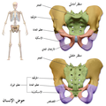
 Bassin osseux-zh.jpg 600 × 600; 94 KB
-
 Bassin osseux.jpg 600 × 600; 60 KB
-
 Bifurcated coccyx & crushed L4.jpg 497 × 800; 83 KB
-
 Bipedalism.jpg 235 × 192; 8 KB
-
 Blausen 0723 Pelvis-ar.png 2,500 × 2,500; 4.41 MB
-
 Blausen 0723 Pelvis.png 2,500 × 2,500; 17.89 MB
-
 BodyParts3D Pelvis.stl 5,120 × 2,880; 711 KB
-
 Braus 1921 231.png 1,424 × 727; 2.97 MB
-
 Bulletin of the Warren Anatomical Museum (1910) (14740147966).jpg 1,708 × 3,172; 1.35 MB
-
 Dem bones (3387445).jpg 1,945 × 1,536; 1.24 MB
-
 Die Frau als Hausärztin (1911) 005 Männliches und weibliches Becken.png 523 × 297; 175 KB
-
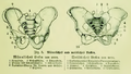
 Die Frau als Hausärztin (1911) 006 Weibliches Becken und seine Teile.png 288 × 358; 123 KB
-
-
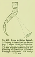 Die Frau als Hausärztin (1911) 183 Grenze des kleinen Beckens.png 248 × 404; 66 KB
-
 Facet Joints Anatomy.jpg 1,018 × 627; 103 KB
-
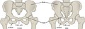 Figure 38 01 12.jpg 1,117 × 387; 193 KB
-
 Grande foro ischiatico.png 514 × 601; 476 KB
-
 Grant 1962 222.png 4,609 × 2,783; 3.9 MB
-
 Grant 1962 223.1.png 1,551 × 2,013; 1.08 MB
-
 Greater sciatic foramen.png 500 × 600; 456 KB
-

-
 Hip bone human.gif 1,928 × 2,000; 84 KB
-
 Hip bone.png 380 × 288; 147 KB
-
 HK TST Science Museum Bones exhibit 32 盆骨 Human pelvis.JPG 3,264 × 2,448; 1.59 MB
-
 Illu pelvic girdle az.jpg 350 × 231; 55 KB
-
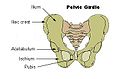 Illu pelvic girdle.jpg 350 × 231; 17 KB
-
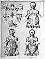 Layers of skin, organs, bones; Vesling "Syntagma", 1647 Wellcome L0007885.jpg 1,194 × 1,564; 822 KB
-
 Lol-bein-mjadmagrind-1.png 605 × 407; 157 KB
-
 Male vs female pelvis LT.PNG 962 × 489; 87 KB
-
 Musée national d'Ethiopie-Au. afarensis-Lucy-Pelvis.jpg 3,591 × 2,493; 2.19 MB
-
 Pelvis (male) 00 - anterior view.png 1,125 × 1,125; 208 KB
-
 Pelvis (male) 00 - lateral view.png 1,125 × 1,125; 128 KB
-
 Pelvis (male) 00 - posterior view.png 1,125 × 1,125; 213 KB
-
 Pelvis (male) 01 - anterior view.png 1,125 × 1,125; 437 KB
-
 Pelvis (male) 01 - lateral view.png 1,125 × 1,125; 251 KB
-
 Pelvis (male) 01 - posterior view.png 1,125 × 1,125; 449 KB
-
 Pelvis (male) 02 - anterior view.png 1,125 × 1,125; 503 KB
-
 Pelvis (male) 02 - lateral view.png 1,125 × 1,125; 264 KB
-
 Pelvis (male) 02 - posterior view.png 1,125 × 1,125; 540 KB
-
 Pelvis (male) 03 - anterior view.png 1,125 × 1,125; 298 KB
-
 Pelvis (male) 03 - inferior view.png 1,125 × 1,125; 263 KB
-
 Pelvis (male) 03 - lateral view.png 1,125 × 1,125; 184 KB
-
 Pelvis (male) 03 - posterior view.png 1,125 × 1,125; 301 KB
-
 Pelvis (male) 03 - superior view.png 1,125 × 1,125; 238 KB
-
 Pelvis (male) animation00.gif 360 × 360; 1.12 MB
-
 Pelvis (male) animation01.gif 360 × 360; 2.31 MB
-
 Pelvis (male) animation02.gif 360 × 360; 3.06 MB
-
 Pelvis (male) animation03.gif 360 × 360; 3.79 MB
-
 Pelvis (PSF).png 2,856 × 1,744; 893 KB
-
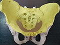
 Pelvis - os coxae, os sacrum (frontal view).jpg 4,608 × 3,456; 4.47 MB
-
 Pelvis - os coxae, os sacrum (lateral view).jpg 3,440 × 2,604; 2.66 MB
-
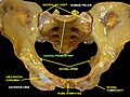
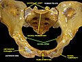
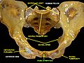
 Pelvis - os coxae, os sacrum (posterior view).jpg 4,140 × 2,712; 3.48 MB
-
 Pelvis - os coxae, os sacrum.jpg 4,608 × 3,456; 4.3 MB
-
 Pelvis 2.jpg 960 × 720; 79 KB
-
 Pelvis CT 3d front 01.jpg 960 × 720; 52 KB
-
 Pelvis diagram es.png 2,277 × 1,704; 111 KB
-
 Pelvis diagram numbered.png 768 × 600; 276 KB
-
 Pelvis diagram zh.png 2,277 × 1,704; 700 KB
-
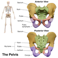
 Pelvis diagram.png 2,277 × 1,704; 704 KB
-
 Pelvis masculina (Elvis). Sima de los Huesos.jpg 4,000 × 2,434; 5.02 MB
-
 Pelvis MH2 Australopithecus sediba.jpg 2,883 × 3,200; 715 KB
-
 Pelvis of E. Welch, showing disease of right hip and disloca Wellcome L0026321.jpg 1,616 × 1,212; 617 KB
-
 Pelvis RG front 01.jpg 960 × 720; 47 KB
-
 Pelvis RG MR CT3d front 01.jpg 417 × 944; 44 KB
-
 Pelvis, from H. Deventer "Operationes Chirurgicae..." Wellcome L0013512.jpg 1,290 × 1,444; 600 KB
-
 Pelvis.jpg 960 × 720; 62 KB
-
 Piştêna hewzê ku.png 1,001 × 493; 240 KB
-
 PSM V22 D339 Pelvic size variations between age and gender.jpg 1,166 × 762; 58 KB
-
 Skeletpelvis-pubis.jpg 1,764 × 1,164; 281 KB
-
 Slide1ab.JPG 960 × 720; 78 KB
-
 Slide1ADAA.JPG 960 × 720; 77 KB
-
 Slide2ab.JPG 960 × 720; 83 KB
-
 Slide3ab.JPG 960 × 720; 83 KB
-
 Slide3ADA-ar.jpg 960 × 720; 162 KB
-
 Slide3ADA.JPG 960 × 720; 81 KB
-
 Slide4ab.JPG 960 × 720; 83 KB
-
 Slide5ab.JPG 960 × 720; 83 KB
-
 Slide6ab.JPG 960 × 720; 84 KB
-
 Slide7ab.JPG 960 × 720; 84 KB
-
 Subpubic angle, female.png 1,226 × 1,019; 529 KB
-
 Subpubic angle, male.png 1,000 × 892; 624 KB
-
 Symphysis Pubis.png 567 × 379; 61 KB
-

-
-
-

-
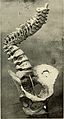
 The principles and practice of obstetrics (1864) (14779931031).jpg 2,608 × 3,446; 1.2 MB
-
-
 UBC-BME-STA-20-007-1.png 1,600 × 866; 72 KB
-
 UBC-BME-STA-20-007-2.png 1,228 × 896; 88 KB
-
 О4о45ор54ор5.tif 934 × 606; 499 KB


_(14581346340).jpg)
.png)


_(14740147966).jpg)
.jpg)
_005_M%C3%A4nnliches_und_weibliches_Becken.png)
_006_Weibliches_Becken_und_seine_Teile.png)
_181_Kn%C3%B6chernes_Becken_mit_Durchmessern.png)
_183_Grenze_des_kleinen_Beckens.png)


_van_licht_in_het_bekken_(tekening_Peter_Kampschuur,_vrij_van_rechten).jpg)


_00_-_anterior_view.png)
_00_-_lateral_view.png)
_00_-_posterior_view.png)
_01_-_anterior_view.png)
_01_-_lateral_view.png)
_01_-_posterior_view.png)
_02_-_anterior_view.png)
_02_-_lateral_view.png)
_02_-_posterior_view.png)
_03_-_anterior_view.png)
_03_-_inferior_view.png)
_03_-_lateral_view.png)
_03_-_posterior_view.png)
_03_-_superior_view.png)
_animation00.gif)
_animation01.gif)
_animation02.gif)
_animation03.gif)
.png)
.jpg)
.jpg)
.jpg)


._Sima_de_los_Huesos.jpg)


_(14590739028).jpg)
_(14774259381).jpg)
_(14777407395).jpg)
_(14777429155).jpg)
_(14779931031).jpg)
_(14758368516).jpg)

