Category:UCSF Chimera
Jump to navigation
Jump to search
software for visualization and analysis of molecular structures  | |||||
| Upload media | |||||
| Instance of | |||||
|---|---|---|---|---|---|
| Has use | |||||
| Developer | |||||
| Sponsor | |||||
| official website | |||||
| |||||
Images of molecules created with the program UCSF Chimera, from University of California, San Francisco.
Media in category "UCSF Chimera"
The following 37 files are in this category, out of 37 total.
-
140210-MGR-CiVSP morph video.ogv 17 s, 640 × 480; 4.98 MB
-
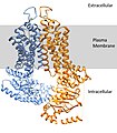 ABCG2 with plasma membrane.jpg 586 × 668; 92 KB
-
 Acetoacetate decarboxylase biounit 3BH3 with inset.png 2,032 × 2,018; 2.41 MB
-
 Active site generated by chimera.jpg 2,241 × 1,138; 539 KB
-
 Adv-D26 7-24-2021 1 ps.tif 10,042 × 8,063; 231.68 MB
-
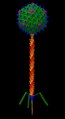
 Bacteriophage Lambda.tif 6,814 × 8,063; 8.11 MB
-
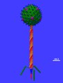
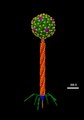
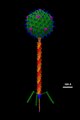
 Bacteriophage phi29.tif 7,618 × 8,063; 14.52 MB
-
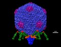
 Bacteriophage T4 Structural Model at Atomic Resolution.tif 9,022 × 8,063; 13.53 MB
-
 Bacteriophage T5 2024 ps.tif 6,130 × 8,063; 7.36 MB
-
 Bacteriophage T5 Structural Model at Atomic Resolution.tif 6,153 × 11,237; 13.74 MB
-
 Bacteriophage T7 7-24-2021 ps.tif 10,366 × 8,063; 239.15 MB
-
 Bacteriophage T7 Structural Model at Atomic Resolution.tif 2,857 × 3,129; 6.21 MB
-
 Benzene-povray.png 564 × 547; 52 KB
-
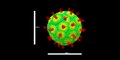
 Bovine Papillomavirus Capsid.png 1,066 × 1,066; 1.97 MB
-
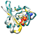
 Catalase Structure.png 3,000 × 3,039; 3.85 MB
-
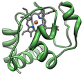 Cytochrome C.png 1,034 × 928; 356 KB
-
 Delphi-peptide-elpot-transparent-3D-sticks.png 1,111 × 787; 124 KB
-
 Final ribbon surface.jpg 1,466 × 724; 133 KB
-
 GAPDH with labels.png 2,048 × 1,217; 1.11 MB
-
 GroEL resolution series.png 2,668 × 742; 1.98 MB
-
 Hras bfactor worm colored by conservation.png 1,300 × 1,236; 691 KB
-
 Hras secondary structure ribbon.png 1,300 × 1,236; 658 KB
-
 Hras surface colored by conservation.png 1,300 × 1,236; 973 KB
-
 Lambda 7-24-2021 ps.tif 5,650 × 8,063; 130.36 MB
-
 Leptin.png 928 × 726; 268 KB
-
 Met-enkephalin 1plx model 1.png 1,600 × 1,218; 317 KB
-
 Mycobacteriophage ZoeJ 7-24-2021 1 ps.tif 4,342 × 8,063; 100.18 MB
-
 Nipah 12142023 1 ps.tif 15,992 × 8,063; 91.35 MB
-
 Pre-mRNA-1ysv-tubes.png 1,111 × 2,048; 965 KB
-
 Protein structure examples for localization.png 1,571 × 1,938; 4.06 MB
-
 Protein structure examples.png 1,571 × 1,938; 3.54 MB
-
 Ribosome shape.png 2,580 × 1,289; 1.72 MB
-
 RNase A.png 3,652 × 2,821; 2.3 MB
-
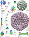
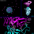 SARS-CoV-2 Infection Structural Analysis.png 7,996 × 8,062; 11.45 MB
-
 SARS-CoV-2 structural model.tiff 7,996 × 4,031; 92.24 MB
-
 Stannabenzene-povray.png 564 × 547; 59 KB
-
 T5 7-24-2021 ps.tif 5,362 × 8,063; 123.71 MB


