Category:RNA structure
Jump to navigation
Jump to search
RNA tertiary (3-dimensional) structure.
Media in category "RNA structure"
The following 61 files are in this category, out of 61 total.
-
12985 2016 561 Fig1A HTML.jpg 567 × 155; 10 KB
-
12985 2016 561 Fig3A HTML.jpg 567 × 296; 16 KB
-
5S ribosomal RNA ribbons.jpg 820 × 768; 169 KB
-
All-atom contacts for two GC basepairs of RNA.jpg 1,031 × 689; 241 KB
-
C6orf10 Stem loop structure.png 388 × 420; 30 KB
-
-
-
DEAD-Box-Helicase-Proteins-Disrupt-RNA-Tertiary-Structure-Through-Helix-Capture-pbio.1001981.s014.ogv 24 s, 512 × 512; 19.41 MB
-
-
-
-
Difference DNA RNA-RU.svg 1,371 × 1,097; 126 KB
-
DNA RNA structure (1).png 1,253 × 1,989; 569 KB
-
DNA RNA structure (2).png 1,253 × 1,989; 541 KB
-
DNA RNA structure (2.1).png 1,253 × 1,989; 584 KB
-
DNA RNA structure (2.2).png 1,253 × 1,989; 559 KB
-
DNA RNA structure (2.3).png 1,253 × 1,989; 568 KB
-
DNA RNA structure (3).png 1,253 × 1,989; 609 KB
-
DNA RNA structure (4).png 1,253 × 1,989; 703 KB
-
DNA RNA structure (full).png 1,253 × 1,989; 798 KB
-
Double-stranded RNA.gif 669 × 669; 3.04 MB
-
G riboswitch RNA ribbon.jpg 760 × 760; 163 KB
-
G riboswitch site w map contacts suite-labels.jpg 1,024 × 768; 540 KB
-
Influenza A Segment 7 Multi-branch Family.jpg 2,477 × 2,342; 874 KB
-
ManA-RNA.svg 311 × 369; 264 KB
-
Ribonucleic acid chemical structure mk.svg 429 × 421; 68 KB
-
Ribonucleic acid chemical structure.svg 429 × 421; 19 KB
-
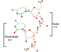
RNA residue-Suite diagram.tif 920 × 780; 793 KB
-
RNA residueSuite diagram.tif 920 × 780; 704 KB
-
RNA structure (1).png 1,247 × 1,989; 530 KB
-
RNA structure (2).png 1,247 × 1,989; 534 KB
-
RNA structure (3).png 1,247 × 1,989; 565 KB
-
RNA structure (4).png 1,246 × 1,989; 610 KB
-
RNA structure (full).png 1,243 × 1,989; 733 KB
-
RNA structuur (1).png 1,608 × 2,492; 1.01 MB
-
RNA structuur (2).png 1,608 × 2,492; 1.01 MB
-
RNA structuur (3).png 1,608 × 2,492; 1.05 MB
-
RNA structuur (4).png 1,608 × 2,492; 1.09 MB
-
RNA structuur (alles).png 1,608 × 2,492; 1.23 MB
-
RNA-Fab crystal.tif 3,840 × 2,160; 10.76 MB
-
Rna-structure.jpg 292 × 351; 10 KB
-
RNASeqPics1.jpg 720 × 540; 46 KB
-
RNASeqPics2.jpg 720 × 540; 33 KB
-
S2m structure of SARS-CoV.png 336 × 805; 53 KB
-
Smotif in RNA suite-labeled.jpg 1,024 × 768; 245 KB
-
TB6Cs1H1 snoRNA.pdf 941 × 941; 4 KB
-
TB6Cs1H3 snoRNA.PNG 420 × 963; 31 KB
-
The-Impact-of-a-Ligand-Binding-on-Strand-Migration-in-the-SAM-I-Riboswitch-pcbi.1003069.s010.ogv 1 min 32 s, 320 × 240; 11.01 MB
-
The-Impact-of-a-Ligand-Binding-on-Strand-Migration-in-the-SAM-I-Riboswitch-pcbi.1003069.s011.ogv 3 min 45 s, 320 × 300; 16.46 MB
-
The-Impact-of-a-Ligand-Binding-on-Strand-Migration-in-the-SAM-I-Riboswitch-pcbi.1003069.s012.ogv 50 s, 794 × 752; 21.07 MB
-
The-Impact-of-a-Ligand-Binding-on-Strand-Migration-in-the-SAM-I-Riboswitch-pcbi.1003069.s013.ogv 59 s, 812 × 750; 35.03 MB
-
The-Impact-of-a-Ligand-Binding-on-Strand-Migration-in-the-SAM-I-Riboswitch-pcbi.1003069.s014.ogv 26 s, 766 × 750; 11.64 MB
-
The-Impact-of-a-Ligand-Binding-on-Strand-Migration-in-the-SAM-I-Riboswitch-pcbi.1003069.s015.ogv 1 min 42 s, 288 × 300; 6.47 MB
-
The-Impact-of-a-Ligand-Binding-on-Strand-Migration-in-the-SAM-I-Riboswitch-pcbi.1003069.s016.ogv 35 s, 768 × 750; 15.95 MB
-
TRNA 1ehz coil lots.jpg 600 × 489; 161 KB
-
Twort groupI intron RNAribbon stereo.jpg 1,024 × 768; 396 KB
-
Varkud satellite ribozyme secondary.png 1,607 × 766; 54 KB
-
Varkud satellite ribozyme tertiary.png 1,632 × 1,043; 604 KB
-
Viruses-11-00298-g002.webp 3,651 × 2,194; 71 KB
-
VS ribozyme dimer 4r4v.png 1,352 × 955; 372 KB
-
YkkC-III-RNA.svg 353 × 115; 170 KB


