Category:Parasitology diagrams
Jump to navigation
Jump to search
Subcategories
This category has the following 2 subcategories, out of 2 total.
Media in category "Parasitology diagrams"
The following 159 files are in this category, out of 159 total.
-
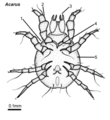
Acarus, female, ventral.png 3,308 × 3,615; 169 KB
-
Aedes thorax parts.png 3,792 × 2,971; 169 KB
-
Amblyomma female dorsal.png 4,561 × 3,436; 189 KB
-
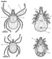
Amblyomma male dorsal ventral.png 4,640 × 2,519; 250 KB
-
Angiostrongylus cantonensis life cycle 01.png 2,899 × 2,313; 570 KB
-
Angiostrongylus costaricensis life cycle.png 2,927 × 2,298; 567 KB
-
Anisakiasis life cycle.png 2,456 × 3,076; 686 KB
-
Anopheles female lateral.png 4,688 × 2,491; 146 KB
-
Argas adult lateral.png 3,920 × 2,785; 183 KB
-
Auchmeromyia larva adult.png 7,078 × 4,700; 362 KB
-
Baylisascaris procyonis life cycle CDC.tif 2,400 × 3,150; 21.65 MB
-
Baylisascaris procyonis life cycle.png 2,453 × 3,162; 774 KB
-
Bovicola chewing louse mouthparts.jpg 1,024 × 581; 96 KB
-
Bovicola female ventral.png 4,174 × 5,574; 296 KB
-
Bovicola louse at skin.png 2,520 × 2,976; 165 KB
-
Brugia malayi life cycle CDC.tif 3,150 × 2,400; 21.65 MB
-
Calliphora larva adult.png 4,906 × 5,015; 432 KB
-
Cephalopina larva.png 5,160 × 1,290; 124 KB
-
Ceratophyllus female lateral.png 3,506 × 2,901; 194 KB
-
Cestode key.pdf 2,100 × 1,275; 666 KB
-
Cheyletiella adult dorsal.png 2,544 × 3,108; 138 KB
-
Chorioptes male female.png 5,236 × 3,952; 291 KB
-
Chrysomya larva adult.png 4,539 × 2,980; 235 KB
-
Chrysomya myiasis skin.png 4,265 × 2,675; 241 KB
-
Cimex female ventral.png 2,815 × 3,740; 205 KB
-
Cochliomyia larva adult.png 4,762 × 2,944; 252 KB
-
Cordylobia larva adult.png 4,671 × 3,469; 255 KB
-
Ctenocephalides adult larva pupa.png 5,252 × 7,620; 489 KB
-
Cuclotogaster female ventral.png 3,475 × 4,822; 247 KB
-
Culex female lateral.png 4,956 × 2,896; 199 KB
-
Culex thorax parts.png 5,034 × 4,438; 327 KB
-
Culicoides female lateral.png 4,924 × 4,123; 387 KB
-
Cuterebra larva adult.png 4,809 × 3,209; 314 KB
-
Cycle Toxoplasma gondii nltxt.jpg 733 × 720; 267 KB
-
Cytodites female male.png 4,876 × 2,524; 206 KB
-
Cytodites mite.jpg 1,298 × 1,536; 1.86 MB
-
Demodex feeding skin.png 2,614 × 3,218; 173 KB
-
Demodex mite ventral.png 2,533 × 5,869; 209 KB
-
Dermacentor female male.png 3,995 × 5,297; 410 KB
-
Dermacentor-female-dorsal.png 3,427 × 2,802; 148 KB
-
Dermacentor-male-dorsal-ventral.png 3,995 × 2,658; 263 KB
-
Dermanyssus female dorsal.png 3,754 × 4,892; 305 KB
-
Dermatobia larvae adult.png 4,431 × 2,739; 215 KB
-
Dermatophagoides female ventral.png 3,524 × 4,346; 237 KB
-
Echidnophaga adult lateral.png 3,133 × 2,822; 164 KB
-
Echinococcus gran LifeCycle lg.jpg 2,000 × 1,562; 521 KB
-
EichlersRule.jpg 546 × 439; 34 KB
-
Enterobius vermicularis life cycle.tif 1,013 × 985; 311 KB
-
Felicola female ventral.png 2,368 × 3,998; 140 KB
-
Gasterophilus larva adult.png 4,788 × 2,852; 303 KB
-
Glossina adult lateral dorsal.png 4,433 × 3,174; 204 KB
-
Glycyphagus female ventral.png 4,082 × 3,945; 212 KB
-
Gnathostoma LifeCycle lg.jpg 1,574 × 2,000; 611 KB
-
Goniocotes female ventral.png 2,646 × 3,452; 159 KB
-
Goniodes female ventral.png 2,646 × 3,835; 183 KB
-
Haemagogus thorax parts.png 7,839 × 4,107; 415 KB
-
Haemaphysalis female male.png 4,015 × 5,796; 400 KB
-
Haemaphysalis-female-dorsal.png 3,724 × 3,217; 184 KB
-
Haemaphysalis-male-dorsal-ventral.png 4,009 × 2,726; 219 KB
-
Haematobia adult lateral.png 4,800 × 3,283; 170 KB
-
Haematopinus female dorsal.png 4,760 × 3,503; 233 KB
-
Haematopota female lateral.png 4,762 × 3,153; 292 KB
-
Heterodoxus female ventral.png 2,646 × 3,448; 166 KB
-
Hippelates adult lateral.png 3,628 × 2,644; 142 KB
-
Hookworm LifeCycle lg.jpg 2,000 × 1,563; 477 KB
-
Hyalomma female male.png 4,399 × 5,947; 464 KB
-
Hyalomma-female-dorsal.png 4,126 × 3,369; 196 KB
-
Hyalomma-male-dorsal-ventral.png 4,399 × 2,759; 271 KB
-
Hybromitra female lateral.png 4,762 × 2,435; 186 KB
-
Hydrotaea adult lateral.png 4,762 × 2,174; 160 KB
-
Hypoderma larva adult.png 4,404 × 3,987; 281 KB
-
Hypoderma larvae skin.png 4,673 × 3,171; 338 KB
-
Ixodes female male.png 4,665 × 5,199; 415 KB
-
Ixodes-female-dorsal-ventral.png 4,654 × 2,993; 244 KB
-
Ixodes-male-dorsal-ventral.png 4,534 × 2,214; 172 KB
-
Knemidokoptes female dorsal ventral.png 4,118 × 2,442; 253 KB
-
KuchTenia19522008 1.jpg 786 × 486; 77 KB
-
KuchTenia19522008 2.jpg 703 × 370; 66 KB
-
Laminosioptes female ventral.png 3,012 × 3,292; 127 KB
-
Leishmaniasis life cycle cdc.tif 3,150 × 2,400; 21.65 MB
-
Lepiselaga-adult-lateral.png 3,640 × 3,336; 221 KB
-
Lepiselaga-adult.tif 3,640 × 3,336; 200 KB
-
Lepiselega female lateral.png 3,840 × 3,420; 223 KB
-
Leptoconops female lateral.png 3,135 × 2,550; 152 KB
-
Linognathus female ventral.png 3,625 × 5,475; 242 KB
-
Linognathus life-cycle.png 6,233 × 5,199; 280 KB
-
Lipeurus male ventral.png 2,061 × 3,622; 138 KB
-
Lucilia larva adult.png 4,696 × 2,999; 227 KB
-
Mansonia thorax parts.png 7,606 × 4,327; 398 KB
-
Margaropus female male.png 3,750 × 4,876; 286 KB
-
Margaropus-female-dorsal.png 3,666 × 3,275; 198 KB
-
Margaropus-male-dorsal.png 2,988 × 1,831; 60 KB
-
Megninia female male.png 5,202 × 3,992; 286 KB
-
Melophagus adult dorsal.png 3,685 × 2,419; 190 KB
-
Melophagus female male puparium.jpg 2,007 × 1,584; 800 KB
-
Menacanthus female ventral.png 3,790 × 4,663; 255 KB
-
Menopon female ventral.png 2,646 × 4,336; 216 KB
-
Multi-host-tick-lifecycle.png 890 × 491; 51 KB
-
Musca feeding skin.png 2,614 × 2,283; 105 KB
-
Myobia mite dorsal.png 4,667 × 3,914; 211 KB
-
Myocoptes female ventral.png 3,780 × 3,153; 155 KB
-
Neotrombicula feeding skin.png 3,483 × 3,704; 247 KB
-
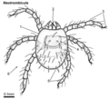
Neotrombicula larva dorsal.png 4,045 × 3,669; 204 KB
-
Notoedres female dorsal ventral.png 4,204 × 2,771; 299 KB
-
Oestrus larva adult.png 4,705 × 4,142; 259 KB
-
Ornithodoros adult lateral.png 3,975 × 2,936; 237 KB
-
Ornithodoros feeding skin.png 3,147 × 2,687; 190 KB
-
Ornithodoros life-cycle pigs.png 4,930 × 2,405; 160 KB
-
Ornithonyssus female dorsal.png 3,840 × 3,228; 209 KB
-
Otobius nymph lateral.png 3,630 × 1,764; 93 KB
-
Otodectes male female revised.png 5,242 × 3,605; 283 KB
-
Otodectes male female.png 5,242 × 3,605; 283 KB
-
Otodectes-mite-copy.png 5,242 × 3,605; 283 KB
-
Pediculus female dorsal.png 3,007 × 3,865; 164 KB
-
Phlebotomus female lateral.png 4,762 × 3,924; 265 KB
-
Phormia larva adult.png 4,792 × 3,020; 248 KB
-
Phormia-larva-adult.png 4,748 × 2,920; 249 KB
-
Plant-parasitic nematode feeding types.jpg 509 × 424; 153 KB
-
Pneumonyssus mite ventral.png 3,912 × 2,549; 160 KB
-
Polyplax-female ventral.png 2,775 × 4,393; 180 KB
-
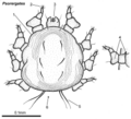
Psorergates female dorsal.png 4,459 × 4,047; 243 KB
-
Psorophora thorax parts.png 7,248 × 4,024; 394 KB
-
Psoroptes feeding skin.png 2,614 × 1,906; 86 KB
-
Psoroptes male female.png 4,992 × 4,488; 286 KB
-
Pthirus female dorsal.png 2,621 × 2,524; 121 KB
-
Pulex adult lateral.png 3,617 × 3,485; 259 KB
-
Quantitative Parasitology 30 windows.jpg 1,116 × 670; 88 KB
-
Quantitative Parasitology on theWeb 10.jpg 921 × 611; 90 KB
-
Rhinoestrus larva adult-head.png 5,291 × 3,123; 229 KB
-
Rhipicephalus female male.png 3,765 × 6,053; 407 KB
-
Rhipicephalus tick life-cycle.png 4,461 × 3,530; 192 KB
-
Rhipicephalus-female-dorsal.png 3,519 × 3,696; 193 KB
-
Rhipicephalus-male-dorsal-ventral.png 3,765 × 2,536; 218 KB
-
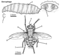
Sarcophaga larva adult.png 5,338 × 4,888; 355 KB
-
Sarcophaga-larva-adult-revised.png 5,338 × 4,888; 355 KB
-
Sarcoptes feeding skin.png 4,376 × 2,604; 237 KB
-
Sarcoptes female dorsal ventral.png 5,226 × 3,603; 308 KB
-
Schistosomiasis life cycle.png 2,803 × 2,393; 825 KB
-
Sites-infestation-mites-skin.png 283 × 219; 69 KB
-
Solenopotes female ventral.png 3,348 × 4,666; 194 KB
-
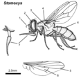
Stomoxys adult lateral.png 2,623 × 2,466; 168 KB
-
Stomoxys-life-cycle.png 5,940 × 3,861; 248 KB
-
Symphoromyia adult lateral.png 4,696 × 2,346; 181 KB
-
Symphoromyia-Adult-Snipe-fly.png 4,608 × 2,248; 180 KB
-
Symphoromyia-Snipe-fly-adult.tif 4,608 × 2,248; 168 KB
-
Toxoplasma gondii Life cycle PHIL 3421 lores hy.png 2,400 × 3,150; 340 KB
-
Toxoplasma LifeCycle CDC.gif 1,130 × 914; 100 KB
-
Triatoma adult dorsal lateral.png 4,094 × 3,460; 289 KB
-
Triatoma assassin-bug.png 4,784 × 1,640; 119 KB
-
Trichodectes female ventral.png 2,603 × 3,590; 186 KB
-
Trichomonas vaginalis LifeCycle CDC.tif 2,400 × 2,912; 20.01 MB
-
Trypanosoma brucei Life Cycle.svg 1,760 × 1,080; 269 KB
-
Trypanosoma cruzi life cycle CDC.tif 3,150 × 2,400; 21.65 MB
-
TrypanosomatidMorphologies Ar.svg 800 × 900; 28 KB
-
TrypanosomatidMorphologies PlainSVG.svg 800 × 900; 27 KB
-
Tunga female.png 4,735 × 2,506; 156 KB
-
Wohlfahrtia larva adult.png 4,134 × 3,552; 244 KB
-
Xenopsylla adult lateral.png 3,632 × 3,205; 238 KB
