Category:Oocytes
Jump to navigation
Jump to search
female gametocyte or germ cell involved early in reproduction; immature precursor of ovum produced in the ovary during female gametogenesis | |||||
| Upload media | |||||
| Instance of | |||||
|---|---|---|---|---|---|
| Subclass of |
| ||||
| Different from | |||||
| |||||
Subcategories
This category has the following 8 subcategories, out of 8 total.
Media in category "Oocytes"
The following 116 files are in this category, out of 116 total.
-
06fertilizado.jpg 434 × 280; 92 KB
-
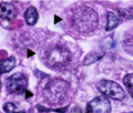
4.B. Ovogonias. Quiescentes en G0-G1 (flechas).png 659 × 559; 600 KB
-
41467 2023 39758 Fig1.webp 1,494 × 1,212; 472 KB
-
41467 2023 39758 Fig2.webp 1,499 × 1,565; 260 KB
-
41467 2023 39758 Fig3.webp 1,500 × 1,525; 326 KB
-
41467 2023 39758 Fig6.webp 1,350 × 1,367; 109 KB
-
A textbook of obstetrics (1899) (14590989838).jpg 2,373 × 1,809; 772 KB
-
-
-
-
-
-
-
-
-
-
-
Abatus cordatus Female and male gametes.jpg 1,172 × 520; 517 KB
-
Aberrant development of in vitro-cultured Prmt5floxflox;Sf1+cre follicles..jpg 1,880 × 2,576; 724 KB
-
Brockhaus and Efron Encyclopedic Dictionary b17 466-0.jpg 777 × 1,118; 164 KB
-
Cdc20-Is-Critical-for-Meiosis-I-and-Fertility-of-Female-Mice-pgen.1001147.s004.ogv 22 s, 1,404 × 700; 527 KB
-
Cdc20-Is-Critical-for-Meiosis-I-and-Fertility-of-Female-Mice-pgen.1001147.s005.ogv 24 s, 1,404 × 700; 616 KB
-
Corona radiata Estructura.png 1,037 × 2,402; 1.4 MB
-
Corona radiata.png 1,182 × 2,397; 3.12 MB
-
Corpusc polar Cortical.png 470 × 895; 514 KB
-
Corpúsculo polar ADN Microtubulos.png 478 × 530; 194 KB
-
Corpúsculo polar Formación.png 358 × 717; 169 KB
-
Corpúsculo polar.png 276 × 245; 40 KB
-
Croissance ovocytaire.png 1,256 × 629; 69 KB
-
Developing-oocytes-and-spermaries-in-Platygyra-acuta.jpg 600 × 255; 116 KB
-
Die Frau als Hausärztin (1911) 148 Menschliche Eizelle.png 138 × 203; 36 KB
-
Egg and Sperm.png 2,000 × 949; 336 KB
-
ESQUEMA-CPEB4.jpg 2,667 × 1,500; 845 KB
-
Essential-Role-for-Endogenous-siRNAs-during-Meiosis-in-Mouse-Oocytes-pgen.1005013.s014.ogv 18 s, 512 × 512; 3.46 MB
-
Foliculo antral.png 766 × 768; 1.04 MB
-
Foliculo de Graaf..png 520 × 358; 441 KB
-
Foliculo FSH.jpg 800 × 397; 126 KB
-
Foliculo maduro.png 770 × 746; 1.24 MB
-
Foliculos ovaricos miguelferig.PNG 516 × 340; 69 KB
-
FOXL2, GATA4, and SMAD3 proteins are expressed in normal follicles and GCTs.png 1,497 × 1,786; 4.68 MB
-
Graafian Follicle Labelled.jpg 1,280 × 720; 157 KB
-
Graafian follicle.jpg 960 × 540; 111 KB
-
Histological and WIHC analysis of Wt1+−; Sf1+− B6 XYB6 fetuses.png 1,234 × 2,748; 4.02 MB
-
ICSI 2 Lainfertilidaddehoy.png 1,059 × 681; 453 KB
-
Immunohistochemical localization of the cell markers in mouse ovaries.png 658 × 1,281; 993 KB
-
Immunoregulation-of-follicular-renewal-selection-POF-and-menopause-in-vivo-vs.-neo-oogenesis-in-1477-7827-10-97-S1.ogv 1 min 15 s, 720 × 240; 4.87 MB
-
-
-
Mbio.00574-21-f001.jpg 1,800 × 1,376; 607 KB
-
Mbio.00574-21-f001a.jpg 486 × 365; 76 KB
-
-
-
-
-
-
-
-
-
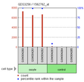
MicroarryOocyte.png 646 × 624; 54 KB
-
Multiple developmental polarities in Drosophila border cell migration.gif 1,000 × 1,123; 138 KB
-
Nucleolos en Ovogonias y Ovocitos. Barra=10µm.png 553 × 256; 167 KB
-
Oocyte Poles.jpg 419 × 653; 37 KB
-
-
LL-Q1860 (eng)-Vealhurl-oosphere.wav 1.2 s; 115 KB
-
Ovocito Cel granulosa cumulus.jpg 683 × 466; 118 KB
-
Ovocito Cuerpo polar.png 350 × 414; 109 KB
-
OVOCITO M2. Polar body 1. DIC microscopy..png 221 × 227; 81 KB
-
Ovocito Meiosis II Asimetría del huso.png 1,924 × 544; 675 KB
-
Ovocito Ovulo Embrion.png 232 × 734; 245 KB
-
Ovocito pre-vitelogenico..png 574 × 330; 187 KB
-
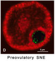
Ovocito preovulatorio Cromatina.png 2,022 × 561; 1.28 MB
-
Ovocito vitelogenico inmaduro. con proyecciones.png 545 × 307; 189 KB
-
Ovocito. Distribuciones de la Telomerasa..png 989 × 2,591; 1.69 MB
-
Ovocitos. Distribución de DNA metiltransferasa3.png 845 × 2,199; 1.52 MB
-
Ovocyte II.png 190 × 190; 214 KB
-
Ovocyte Mr HAMAMAH.jpg 1,004 × 440; 62 KB
-
Ovulo Embrion.png 526 × 534; 205 KB
-
Pig oocyte dapi 1.jpg 2,560 × 1,920; 1.39 MB
-
Pig oocyte dapi 2.jpg 2,560 × 1,920; 1.45 MB
-
Pig oocyte dapi 3.jpg 2,560 × 1,920; 1.85 MB
-
Pig oocyte dapi 4.jpg 2,560 × 1,920; 1.74 MB
-
Prefertilization Mammalian Egg Cell.PNG 512 × 384; 10 KB
-
Prmt5 was deleted in granulosa cells of Prmt5floxflox;Sf1+cre mice.jpg 1,654 × 1,529; 627 KB
-
PRMT5 was expressed in granulosa cells of growing follicles.jpg 2,528 × 2,120; 959 KB
-
-
-
-
-
-
-
Shape change of a starfish oocyte during meiotic cell division.png 1,826 × 922; 1.52 MB
-
Steroidogenesis in theca and granulosa cells.png 2,053 × 1,058; 1.47 MB
-
Strange mitotic oocytes of primary follicles.png 4,069 × 3,051; 8.94 MB
-
The-Biphasic-Increase-of-PIP2-in-the-Fertilized-Eggs-of-Starfish-New-Roles-in-Actin-Polymerization-pone.0014100.s003.ogv 9.3 s, 1,024 × 1,024; 5.72 MB
-
-


