Category:Non-coding RNA
Jump to navigation
Jump to search
class of RNA that is not translated into proteins | |||||
| Upload media | |||||
| Subclass of | |||||
|---|---|---|---|---|---|
| |||||
This category groups pages related to non-coding RNA, which is cellular ribonucleic acid (RNA) that is not translated into a protein but fulfills another role.
Subcategories
This category has the following 9 subcategories, out of 9 total.
A
- Antitoxin HigA (2 F)
B
- Bacterial small RNA (31 F)
H
- Hairpin ribozyme (5 F)
L
- Long non-coding RNAs (7 F)
R
T
X
- Xist (14 F)
Pages in category "Non-coding RNA"
This category contains only the following page.
Media in category "Non-coding RNA"
The following 200 files are in this category, out of 1,288 total.
(previous page) (next page)-
23S methyl RNA motif secondary structure.jpg 452 × 452; 11 KB
-
4rfam scaRNA12 U89 stk.svg 331 × 621; 248 KB
-
4rfam scaRNA13 stk.svg 415 × 224; 253 KB
-
4rfam scaRNA16 stk.svg 239 × 287; 199 KB
-
4rfam scaRNA5 stk.svg 202 × 637; 255 KB
-
4rfam scaRNA6 stk.svg 318 × 555; 249 KB
-
4rfam scaRNA7 stk.svg 275 × 575; 232 KB
-
4rfam scaRNA9 stk.svg 382 × 231; 160 KB
-
4rfam snora53 stk.svg 207 × 419; 232 KB
-
4rfam snord17 stk.svg 420 × 441; 177 KB
-
4rfam snord22 stk.svg 308 × 340; 157 KB
-
4rfam U6-77 stk.svg 344 × 383; 165 KB
-
4rfam U85 stk.svg 301 × 794; 277 KB
-
-
-
A-MAPK-Driven-Feedback-Loop-Suppresses-Rac-Activity-to-Promote-RhoA-Driven-Cancer-Cell-Invasion-pcbi.1004909.s011.ogv 2.9 s, 1,024 × 1,024; 1.09 MB
-
-
-
-
ACAT SS.png 400 × 460; 48 KB
-
-
AdoCbl riboswitch secondary structure.jpg 452 × 520; 41 KB
-
AHBV epsilon secondary structure.jpg 452 × 520; 43 KB
-
ALIL pk.png 400 × 460; 42 KB
-
Alirna.svg 558 × 99; 57 KB
-
ATPC secondary structure.jpg 452 × 452; 67 KB
-
B55 SScons.png 603 × 693; 31 KB
-
Bacteroid-trp SS.png 400 × 460; 32 KB
-
-
Bxd c1 SS.png 400 × 460; 40 KB
-
BYDV SS.png 400 × 460; 87 KB
-
BZIPintron animal.svg 512 × 419; 17 KB
-
BZIPintron ascomycota.svg 512 × 920; 19 KB
-
BZIPintron basidiomycota.svg 512 × 530; 22 KB
-
BZIPintron candida.svg 512 × 1,197; 86 KB
-
BZIPintron plant.svg 512 × 490; 26 KB
-
BZIPintron saccharomycetales.svg 512 × 1,179; 20 KB
-
C-di-GMP.svg 711 × 441; 27 KB
-
CDKN2B-AS secondary structure.jpg 452 × 520; 40 KB
-
CeN115 secondary structure.jpg 452 × 520; 49 KB
-
CeN23-1 secondary structure.jpg 452 × 520; 42 KB
-
CeN56 secondary structure.jpg 452 × 520; 53 KB
-
CeN93 secondary structure.jpg 452 × 520; 26 KB
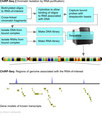
-
ChiRP-Seq.png 497 × 584; 69 KB
-
CIIRNA.png 603 × 693; 48 KB
-
Class I Ligase Ribozyme.jpg 894 × 976; 433 KB
-
Codon-Anticodon pairing.svg 620 × 608; 20 KB
-
CrfA SScons.png 603 × 693; 51 KB
-
Csfg.png 1,479 × 910; 156 KB
-
CspA 5'UTR.png 603 × 693; 66 KB
-
Cyclic di-GMP riboswitch secondary structure.jpg 452 × 452; 17 KB
-
DNA to protein or ncRNA.svg 1,200 × 824; 1.54 MB
-
-
-
-
-
-
-
Drz-agam-1 SS cons.png 603 × 693; 48 KB
-
Drz-agam-1-3 SScons.png 603 × 693; 58 KB
-
Drz-agam-2-2 SScons.png 603 × 693; 43 KB
-
Drz-Bflo-2 SScons.png 603 × 693; 56 KB
-
Drz-Cjap-1 SScons.png 603 × 693; 43 KB
-
Drz-Spur-1 SScons.png 603 × 693; 54 KB
-
-
-
-
-
EBER SScons.png 603 × 693; 59 KB
-
EBER2 Figure.jpg 1,547 × 725; 110 KB
-
-
-
-
-
-
Endothelial-Cells-Use-a-Formin-Dependent-Phagocytosis-Like-Process-to-Internalize-the-Bacterium-ppat.1005603.s010.ogv 6.3 s, 1,024 × 1,024; 20.39 MB
-
-
-
ENOD40 secondary structure and sequence conservation.png 400 × 460; 57 KB
-
ESQUEMA.pdf 1,239 × 1,752; 176 KB
-
Evf2-ss-cons.png 400 × 460; 35 KB
-
Fgene-03-00132-g002 1.jpg 867 × 962; 483 KB
-
Flmb SScons.png 603 × 693; 56 KB
-
FnrS SScons.png 400 × 460; 47 KB
-
FourU.png 833 × 958; 151 KB
-
FstAT SScons.png 603 × 693; 65 KB
-
GABRA3RNA.png 400 × 400; 37 KB
-
Gadd7 ss.png 603 × 693; 32 KB
-
GIR1 SS.png 400 × 460; 92 KB
-
GP knot1 secondary structure.jpg 452 × 452; 59 KB
-
GP knot2 secondary structure.jpg 452 × 452; 59 KB
-
GRIK4 3p UTR secondary structure.jpg 452 × 452; 42 KB
-
HIV-1 SD secondary structure.jpg 452 × 520; 68 KB
-
HIV-1 SL3 secondary structure.jpg 452 × 520; 69 KB
-
HIV-1 SL4 secondary structure.jpg 452 × 520; 69 KB
-
HOTAIR 1 secondary structure.jpg 452 × 520; 78 KB
-
HOTAIR 2 secondary structure.jpg 452 × 520; 42 KB
-
HOTAIR 3 secondary structure.jpg 452 × 520; 56 KB
-
HOTAIR 4 secondary structure.jpg 452 × 520; 34 KB
-
HOTAIR 5 secondary structure.jpg 452 × 520; 38 KB
-
HSR-omega SS.png 400 × 460; 40 KB
-
HSUR1 SScons.png 603 × 693; 37 KB
-
HSUR2 SScons.png 603 × 693; 41 KB
-
IbpB Thermometer.png 400 × 460; 67 KB
-
InvR secondary structure.jpg 452 × 452; 47 KB
-
IscRS SS.png 400 × 460; 53 KB
-
IstR SScons.png 400 × 460; 38 KB
-
KCNQ1DN secondary structure.jpg 452 × 520; 56 KB
-
KIF5B-and-Nup358-Cooperatively-Mediate-the-Nuclear-Import-of-HIV-1-during-Infection-ppat.1005700.s006.ogv 46 s, 1,346 × 815; 1.23 MB
-
KIF5B-and-Nup358-Cooperatively-Mediate-the-Nuclear-Import-of-HIV-1-during-Infection-ppat.1005700.s007.ogv 14 s, 1,648 × 1,002; 465 KB
-
MALAT1 secondary structure.jpg 452 × 520; 54 KB
-
Mature mir-155-5p and -3p.png 902 × 187; 41 KB
-
MFR secondary structure.jpg 452 × 520; 53 KB
-
MgrR SScons.png 603 × 693; 50 KB
-
MGsensor SS.png 400 × 460; 39 KB
-
MicroRNA-34 precursor structure.svg 217 × 575; 87 KB
-
-
-
-
-
MicX SScons.png 603 × 693; 54 KB
-
Mini-ykkC secondary structure.jpg 452 × 452; 46 KB
-
MiR-10 consensus structure.jpg 452 × 933; 213 KB
-
Mir-134 SS.png 400 × 460; 34 KB
-
Mir-138 SS.png 400 × 460; 34 KB
-
Mir-144 SS.png 400 × 460; 33 KB
-
Mir-146 SS.png 400 × 460; 38 KB
-
Mir-150 SS.png 400 × 460; 30 KB
-
Mir-191 SS.png 400 × 460; 29 KB
-
Mir-207 SS.png 400 × 460; 32 KB
-
Mir-208 SS.png 400 × 460; 33 KB
-
Mir-214 SS.png 400 × 460; 31 KB
-
Mir-224 SS.png 400 × 460; 30 KB
-
Mir-27 SS.png 400 × 460; 39 KB
-
Mir-296 SS.png 400 × 460; 30 KB
-
Mir-33 SS.png 400 × 460; 37 KB
-
Mir-338 SS.png 400 × 460; 37 KB
-
Mir155 gene.png 741 × 270; 27 KB
-
MirR-155 and AGT1R.png 1,038 × 210; 51 KB
-
MOCO RNA motif secondary structure.jpg 452 × 452; 54 KB
-
NcRNA biogenesis (vector).svg 1,067 × 837; 89 KB
-
NcRNAs-central-dogma.svg 837 × 436; 16 KB
-
NEAT1 paraspeckles in U-2 OS cells.jpg 937 × 938; 224 KB
-
NRON secondary structure.jpg 452 × 452; 37 KB
-
OLE secondary structure.jpg 452 × 452; 22 KB
-
OmrA-B RNA structure.png 460 × 615; 74 KB
-
OmrA-B RNA structure.svg 460 × 614; 80 KB
-
PabO44.jpg 858 × 523; 56 KB
-
PCA3 1 secondary structure.jpg 452 × 520; 45 KB
-
PCA3 2 secondary structure.jpg 452 × 520; 68 KB
-
PCGEM1 secondary structure.jpg 452 × 520; 58 KB
-
PHYLIS SS.png 400 × 460; 36 KB
-
PiRNA.jpg 509 × 106; 5 KB
-
PK-G12rRNA secondary structure.jpg 452 × 452; 19 KB
-
Pre-mir-155.png 710 × 180; 5 KB
-
PreQ1-II secondary structure.jpg 452 × 452; 67 KB
-
PRNA SS.png 400 × 460; 36 KB
-
Pseudomonas Rsm.png 555 × 596; 97 KB
-
PtaRNA phylogenetic tree.svg 702 × 954; 25 KB
-
PtaRNA1 SS.png 400 × 460; 34 KB
-
PurD secondary structure.jpg 452 × 452; 30 KB
-
Pxr.png 603 × 693; 26 KB
-
-
Rabies-Virus-Infection-Induces-the-Formation-of-Stress-Granules-Closely-Connected-to-the-Viral-ppat.1005942.s002.ogv 9.9 s, 1,352 × 694; 1,019 KB
-
RatA SScons.png 603 × 693; 52 KB
-
RdlD SScons.png 603 × 693; 47 KB
-
Revcen SScons.png 603 × 693; 48 KB
-
RF site1 secondary structure.jpg 452 × 452; 56 KB
-
RF site2 secondary structure.jpg 452 × 452; 46 KB
-
RF site3 secondary structure.jpg 452 × 452; 60 KB
-
RF site4 secondary structure.jpg 452 × 452; 48 KB
-
RF site5 secondary structure.jpg 452 × 452; 44 KB
-
RF site6 secondary structure.jpg 452 × 452; 38 KB
-
RF site8 secondary structure.jpg 452 × 452; 52 KB
-
RF site9 secondary structure.jpg 452 × 452; 54 KB
-
RF00001.jpg 452 × 452; 16 KB
-
RF00002.jpg 452 × 452; 23 KB
-
RF00003-rscape.svg 276 × 204; 82 KB
-
RF00003.jpg 452 × 452; 20 KB
-
RF00004.jpg 452 × 452; 34 KB
-
RF00005.jpg 609 × 609; 40 KB
-
RF00006.jpg 609 × 609; 26 KB
-
RF00007.jpg 452 × 452; 15 KB
-
RF00008-rscape.svg 146 × 126; 27 KB
-
RF00008.jpg 609 × 609; 37 KB
-
RF00009.jpg 452 × 452; 18 KB
-
RF00010.jpg 452 × 452; 22 KB
-
RF00011.jpg 800 × 798; 86 KB
-
RF00012.jpg 452 × 452; 16 KB
-
RF00013-rscape.svg 108 × 616; 93 KB
-
RF00013.jpg 452 × 452; 10 KB
-
RF00014.jpg 452 × 452; 21 KB
-
RF00015.jpg 452 × 452; 15 KB
-
RF00016.jpg 452 × 452; 15 KB
-
RF00017 hsap.jpg 1,337 × 825; 164 KB
-
RF00017.jpg 800 × 798; 32 KB
-
RF00018.jpg 452 × 452; 17 KB
-
RF00019.jpg 452 × 452; 13 KB
-
RF00020.jpg 452 × 452; 15 KB


