Category:Microscopy staining methods
Jump to navigation
Jump to search
technique used to enhance contrast of specimens observed under a microscope | |||||
| Upload media | |||||
| Instance of |
| ||||
|---|---|---|---|---|---|
| Subclass of |
| ||||
| Part of | |||||
| Different from | |||||
| |||||
Subcategories
This category has the following 34 subcategories, out of 34 total.
B
- Bowdian stains (2 F)
C
- Cell staining (33 F)
- Chlorazol black stains (1 F)
- Cytoplasmic staining (6 F)
D
E
- EdU (4 F)
F
G
- Gentian violet stains (2 F)
- Gomori trichrome stain (4 F)
H
- H&E stain (81 F)
I
- India ink stains (6 F)
L
M
- Masson's trichrome stain (6 F)
- Methylene blue stains (22 F)
- Mucicarmine stains (2 F)
N
- Nissl stain (17 F)
- Nuclear staining (23 F)
O
- Orthochromasia (43 F)
P
- Pappenheim's stain (5 F)
- Periodic acid–Schiff stain (32 F)
R
- Reticulin stain (9 F)
S
- Silver staining (26 F)
T
- Trypan blue staining (10 F)
V
- Van Gieson's stain (1 F)
W
- Wright's stain (10 F)
Z
- Ziehl-Neelsen stain (19 F)
Media in category "Microscopy staining methods"
The following 60 files are in this category, out of 60 total.
-
2017 1101 HorseEos 40x 6-crop-wiki2.tif 640 × 640; 1.17 MB
-
Albumen stained.jpg 756 × 572; 303 KB
-
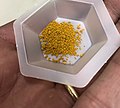
Alizarin Red S powder in boat.jpg 2,500 × 2,253; 1.13 MB
-
Alizarin Red S powder.jpg 1,705 × 1,306; 1.08 MB
-
Anal sac adenocarcinoma cytology.JPG 1,834 × 1,297; 337 KB
-
Asparagus parenchyma& vascular bundle.jpg 1,146 × 872; 487 KB
-
Asparagus vascular bundle.jpg 1,146 × 872; 354 KB
-
Bacteria on Warthin–Starry stain.jpg 983 × 734; 810 KB
-
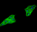
Breast cancer cells.tif 512 × 512; 770 KB
-
C4orf50 stain.png 428 × 428; 351 KB
-
Canine histiocytoma cytology.JPG 1,503 × 949; 206 KB
-
Canine lymphoma 1.JPG 1,192 × 885; 124 KB
-
Canine transmissible venereal tumor cytology.JPG 1,553 × 1,079; 194 KB
-
Cardiac Stem Cell Differentiation.png 512 × 444; 64 KB
-
Chlamydia Geimsa Stain CDC.jpg 699 × 456; 113 KB
-
CongoRed.JPG 990 × 887; 481 KB
-
Deroceras laeve epithelium 400 saturn.jpg 768 × 1,024; 246 KB
-
Deroceras laeve hepatopancreas 400 saturn.jpg 1,024 × 768; 208 KB
-
Deroceras laeve muscle 200 HE.jpg 1,024 × 768; 242 KB
-
Deroceras laeve muscle 400 orcein.jpg 1,024 × 768; 270 KB
-
Deroceras laeve radula 400 orcein.jpg 1,024 × 768; 218 KB
-
Differentiated 3T3-L1 Cell line stained with Oil O Red.jpg 398 × 344; 57 KB
-
Dorsal root ganglion on polypyrrole fibers.tif 1,149 × 525; 476 KB
-
Genesis of nematocysts.JPG 4,000 × 3,000; 3.88 MB
-
Giardia cyst.JPG 1,192 × 803; 63 KB
-
Glass Slide.jpg 3,968 × 2,976; 2.89 MB
-
Glass stained histologic sections.jpg 2,560 × 1,920; 825 KB
-
Gram positive cocci in singles, pairs and chains.jpg 4,000 × 2,250; 1.19 MB
-
Human metaphase painted with mouse chromosome 11 probe.png 896 × 871; 341 KB
-
Ileon-Payer-AZAN.jpg 1,800 × 1,447; 771 KB
-
Isorhiza nematocyst.tif 1,600 × 1,200; 5.49 MB
-
Laser capture microdissection and four staining methods.jpg 900 × 1,203; 373 KB
-
Marburg virus liver injury.jpg 1,756 × 1,224; 403 KB
-
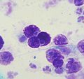
Mast cell tumor cytology 1.JPG 1,201 × 873; 116 KB
-
Mast cell tumor cytology 2.JPG 1,209 × 1,143; 183 KB
-
Melanoma - cytology field stain.jpg 3,324 × 2,348; 3.05 MB
-
Microsporum canis 1.JPG 1,198 × 876; 78 KB
-
Monocotyledon structure.jpg 565 × 519; 212 KB
-
Mouth cells.jpg 400 × 300; 74 KB
-
NSMT-PL 570 7.18 mm frontal.jpg 1,242 × 846; 176 KB
-
Olive infiorescence.jpg 1,840 × 1,232; 766 KB
-
Parasite170078-fig2 Cichlidogyrus philander (Monogenea, Ancyrocephalidae) (main image).png 2,107 × 1,035; 3.06 MB
-
Parasite170078-fig2 Cichlidogyrus philander (Monogenea, Ancyrocephalidae).png 2,120 × 2,120; 6.46 MB
-
Perianal gland tumor cytology.JPG 1,385 × 979; 123 KB
-
PHIL 2767 Poliovirus Myotonic dystrophic changes.jpg 2,932 × 1,972; 843 KB
-
Plant microscopy2.jpg 3,010 × 3,023; 866 KB
-
Plenty of pus cells in Urine Microscopy.jpg 4,000 × 2,250; 2.42 MB
-
Pluteus.cervinus.cystidia.400x.JPG 1,488 × 1,793; 750 KB
-
RBCs and Yeasts in Urine Microscopy.jpg 4,000 × 3,000; 1.16 MB
-
SHG image of elastin in liver.jpg 636 × 690; 96 KB
-
Sperm viability test.jpg 2,533 × 2,322; 655 KB
-
Spermatozoa in Ziehl-Neelsen stained smear of Semen.jpg 4,000 × 2,250; 1.87 MB
-
Sperms in Giemsa Stained smear of Semen.jpg 4,000 × 3,000; 1.49 MB
-
Striatal Medium-Sized Spiny Neuron.jpg 1,024 × 1,024; 88 KB
-
Tobacco Mosaic Virus.jpg 524 × 500; 52 KB
-
Veins of Resilience.jpg 4,888 × 4,944; 25.03 MB
-
Xanthomonas Campestris under the miscroscope.jpg 958 × 1,280; 153 KB
-
Сорус папоротника Polypodium aureum 2.jpg 3,543 × 2,418; 7.54 MB
-
Тимус.jpg 18,229 × 18,229; 227.83 MB


