Category:Gram stains
Jump to navigation
Jump to search
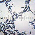
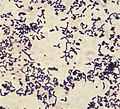
English: Gram staining (or Gram's method) is an empirical method of differentiating bacterial species into two large groups (Gram-positive and Gram-negative) based on the chemical and physical properties of their cell walls.
The method is named after its inventor, the Danish scientist Hans Christian Gram (1853 – 1938), who developed the technique in 1884 to discriminate between pneumococci and Klebsiella pneumoniae bacteria.
Deutsch: Gram-Färbung ist eine empirische Methode zur Unterscheidung von Bakterienarten in zwei große Gruppen (Gram-positive und Gram-negative) auf der Grundlage der chemischen und physikalischen Eigenschaften ihrer Zellwände. Durch das Färbeverfahren erscheinen im Lichtmikroskop grampositive Bakterien blau, gramnegative rot. Die Methode ist nach ihrem Erfinder, dem dänischen Wissenschaftler Hans Christian Gram (1853 - 1938) benannt, der 1884 die Technik entwickelte, um zwischen Pneumokokken und Klebsiella pneumoniae Bakterien zu unterscheiden.
microbiological method for identification; method of staining used to differentiate bacterial species into two large groups (gram-positive and gram-negative) | |||||
| Upload media | |||||
| Instance of |
| ||||
|---|---|---|---|---|---|
| Subclass of | |||||
| Named after | |||||
| Discoverer or inventor | |||||
| |||||
Subcategories
This category has the following 4 subcategories, out of 4 total.
G
S
Media in category "Gram stains"
The following 130 files are in this category, out of 130 total.
-
11G0002 lores.jpg 700 × 470; 78 KB
-
20100905 205851 Streptobacilli.jpg 1,600 × 989; 129 KB
-
20100905 211652 SpirochetesZoom.jpg 1,577 × 1,146; 369 KB
-
20101017 175758 Bacilli.jpg 3,072 × 2,304; 290 KB
-
20101017 175839 BacilliFromSqwincher.jpg 1,600 × 1,600; 193 KB
-
20101017 231210 Staphylococcus.jpg 1,500 × 1,500; 221 KB
-
20101210 020132 StreptococcusThermophilus.jpg 1,400 × 1,000; 78 KB
-
20101212 205549 LactobacillusAcidophilus.jpg 3,072 × 2,304; 327 KB
-
20110322 193829 Bacteria SMB UTI.jpg 1,600 × 1,600; 258 KB
-
Aerococcus urinae - microscopy.jpg 1,920 × 1,080; 707 KB
-
Aeromonas veronii biovar sobria Gram Stain on Microscope Slide.jpg 1,796 × 1,796; 364 KB
-
Bacillus anthracis Gram.jpg 2,892 × 1,940; 6.46 MB
-
Bacillus cereus Gram.jpg 600 × 450; 17 KB
-
Bacillus coagulans 01.jpg 700 × 460; 56 KB
-
Bacillus subtilis gram stain CDC PHIL 19261.jpg 700 × 460; 19 KB
-
Bacillus subtilis Gram stain.jpg 2,080 × 1,536; 2.31 MB
-
Bacillus subtilis Gram.jpg 500 × 375; 38 KB
-
Bacterial cell wall.png 600 × 405; 105 KB
-
Bacterial vaginosis Gram stain.jpg 569 × 804; 828 KB
-
BacteroidesFragilis Gram.jpg 700 × 468; 48 KB
-
Bifidobacterium adolescentis Gram.jpg 600 × 450; 35 KB
-
Bordetella pertussis.jpg 2,588 × 2,383; 2.15 MB
-
Brucella melitensis.jpg 2,880 × 2,185; 1.06 MB
-
Brucella spp.JPG 2,835 × 2,257; 2.42 MB
-
C tetani gram stain.jpg 440 × 298; 21 KB
-
Candida dubliniensis.jpg 1,600 × 1,200; 586 KB
-
Citrobacter freundii Gram stain.jpg 2,080 × 1,536; 2.2 MB
-
Clostridi bacteria gram coloration.jpg 2,425 × 2,424; 1.63 MB
-
Clostridium perfringens gas gangrene.jpg 1,200 × 890; 198 KB
-
Clostridium perfringens.jpg 1,813 × 1,206; 2.64 MB
-
Corynebacterium xerosis Gram stain.jpg 720 × 542; 72 KB
-
Diphtheroides.jpg 1,912 × 1,108; 936 KB
-
Diphtheroids as pathogens in Gram staining of sputum.jpg 4,000 × 2,250; 1.06 MB
-
E choli Gram.JPG 1,609 × 1,461; 872 KB
-
E.coli gram stain.jpg 4,032 × 3,024; 1.88 MB
-
Encapsulated strain of Streptococcus pneumoniae in clinical sample sputum Gram staining.jpg 4,160 × 2,340; 2.82 MB
-
Enterobacter aerogenes.jpg 2,080 × 1,536; 2.19 MB
-
Enterococcus histological pneumonia 01.png 597 × 434; 416 KB
-
Escherichia coli Gram.jpg 500 × 375; 36 KB
-
Gasbrand02.JPG 1,600 × 1,200; 184 KB
-
Gonococcal urethritis PHIL 4085 lores.jpg 3,604 × 2,376; 3.5 MB
-
Gram - algorithm.png 1,089 × 518; 35 KB
-
Gram -Positive and Negative Bacteria.jpg 4,000 × 2,250; 1.16 MB
-
Gram escherichia coli und micrococcus luteus.jpg 387 × 290; 38 KB
-
Gram Negative Rods in Sputum Gram Staining.jpg 4,000 × 2,250; 1 MB
-
Gram Negative Rods of Aeromonas hydrophila.jpg 4,000 × 2,250; 1.08 MB
-
Gram Negative Rods of Serratia fonticola.jpg 4,000 × 2,250; 1.46 MB
-
Gram Positive Classification.png 667 × 458; 69 KB
-
Gram positive cocci in chains of Streptococcus agalactiae.jpg 4,000 × 2,250; 1.4 MB
-
Gram positive yeast cells in Gram staining of culture.jpg 1,920 × 1,080; 288 KB
-
Gram Stain Anthrax.jpg 600 × 405; 41 KB
-
Gram stain of Rothia dentocariosa.jpg 1,112 × 1,009; 257 KB
-
Gram stain of Streptococcus pneumoniae.jpg 2,980 × 2,322; 1.38 MB
-
Gram stain saliva.jpg 1,179 × 657; 678 KB
-
Gram Stain.png 1,280 × 720; 221 KB
-
Gram Staining Process.jpg 4,032 × 3,024; 2.37 MB
-
Gram variability.jpg 939 × 492; 249 KB
-
Gram- negative bacteria.jpg 452 × 604; 94 KB
-
Gram-negative Bacteria - Lab methods algorithm.svg 1,350 × 600; 16 KB
-
Gram-positive stain.jpg 800 × 533; 83 KB
-
Gram-Stained Yogurt Bacteria.jpg 9,000 × 12,000; 6.49 MB
-
Gram-staining-of-D.jpg 600 × 449; 98 KB
-
Haemophilus influenzae Gram.JPG 1,902 × 1,591; 1.15 MB
-
Haemophilus influenzae sputum 1000x edited.jpg 1,920 × 1,200; 915 KB
-
Ideal smear of Sputum.jpg 4,000 × 3,000; 2.64 MB
-
Klebsiella oxytoca.jpg 2,080 × 1,536; 2.19 MB
-
Lactobacilli (Gram stain).jpg 3,008 × 2,000; 2.13 MB
-
Lactobacillus sp 01.png 625 × 498; 257 KB
-
LegionellaPneumophila Gram.jpg 700 × 475; 54 KB
-
Leuconostoc mesenteroides Gram Staining.jpg 4,000 × 3,000; 1.32 MB
-
LF and NLF Gram Negative Bacteria on MacConkey medium.jpg 2,340 × 4,160; 2.83 MB
-
Methylobacterium (Gram stain).jpg 2,080 × 1,536; 2.09 MB
-
Microbiology gram stain.jpg 4,032 × 3,024; 2.31 MB
-
Neisseria gonorrhoeae and pus cells.jpg 3,604 × 2,925; 1.09 MB
-
Neisseria gonorrhoeae diplococci inside a neutrophil.jpg 2,232 × 1,645; 357 KB
-
Neisseria gonorrhoeae PHIL 3693 lores.jpg 1,774 × 1,158; 517 KB
-
Neisseria gonorrhoeae.jpg 5,312 × 2,988; 2.85 MB
-
Neisseria meningitidis CSF Gram 1000.jpg 1,920 × 1,200; 639 KB
-
Nocardia in Gram Stain.tif 1,360 × 1,024; 3.99 MB
-
Nocardiosis - Gram stain Case 149 (5286067518).jpg 1,280 × 960; 646 KB
-
Nocardiosis - Gram stain Case 149 (5286067644).jpg 1,280 × 960; 661 KB
-
Non-Ideal smear of Sputum.jpg 4,000 × 3,000; 2.3 MB
-
Normal flora, Pus cells and Epithelial cells.jpg 4,000 × 3,000; 1.39 MB
-
Numerous Gram Negative Bacteria and Pus cells in Gram staining of sputum.jpg 4,000 × 2,250; 1,020 KB
-
Plenty of pus cells and Gram negative diplococci in Gram stained smear of sputum.jpg 4,160 × 2,340; 1.46 MB
-
Pseudomonas aeruginosa Gram.jpg 500 × 375; 66 KB
-
Pseudomonas aeruginosa gram.jpg 590 × 499; 50 KB
-
Pseudomonas aeruginosa smear Gram 2010-02-10.JPG 1,342 × 1,006; 649 KB
-
Pseudomonas fluorescens Gram Stain on Microscope Slide.jpg 1,200 × 1,200; 454 KB
-
Pseudomonas fluorescens.jpg 2,080 × 1,536; 2.35 MB
-
Purulent inflammation, Gram stain 3.jpg 1,920 × 1,280; 1.47 MB
-
Pus cells with Neisseria gonorrhoeae.jpg 3,008 × 2,000; 2.13 MB
-
Pus cells.jpg 4,000 × 3,000; 563 KB
-
Rhodococcus fascians.jpg 2,080 × 1,536; 2.21 MB
-
Rothia dentocariosa PHIL15195.png 3,045 × 2,005; 13.29 MB
-
Rothia dentocariosa PHIL21290.png 3,045 × 2,005; 12.36 MB
-
Rothia dentocariosa PHIL21292.png 3,045 × 2,005; 14.64 MB
-
Rothia dentocariosa PHIL21293.png 3,045 × 2,005; 15.7 MB
-
Rothia dentocariosa PHIL21294.png 3,045 × 2,005; 14.31 MB
-
Salmonella Typhimurium Gram.jpg 500 × 375; 84 KB
-
Shigella flexneri Gram Stain on Microscope Slide.jpg 1,220 × 1,220; 469 KB
-
Shigella flexneri Gram.jpg 500 × 375; 30 KB
-
ShIgella sonnei Gram negative rods.jpg 4,000 × 3,000; 1.36 MB
-
Spermatozoa in Gram Stained Smear of Semen.jpg 4,000 × 2,250; 1.04 MB
-
Staph sputum.JPG 1,584 × 1,523; 929 KB
-
Staphylococcus aureus Gram.jpg 500 × 375; 24 KB
-
Staphylococcus saprophyticus.jpg 2,080 × 1,536; 2.3 MB
-
Stenotrophomonas maltophilia.jpg 2,080 × 1,536; 2.14 MB
-
Streptobacilli and streptococci in Gram stained smear microscopy at 1000X magnification.jpg 4,160 × 2,340; 1.23 MB
-
Streptococcus mutans 01.jpg 658 × 481; 87 KB
-
Streptococcus mutans Gram.jpg 600 × 450; 41 KB
-
StreptococcusMutans.jpg 654 × 473; 63 KB
-
The Gram Staining - Bacteria Gram Negative.JPG 2,448 × 3,264; 1.92 MB
-
Tincion diferencial.png 209 × 291; 136 KB
-
Vibrio cholerae gram stain CDC.jpg 700 × 723; 56 KB
-
Yeast cells and short hyphae in Gram stain.jpg 4,000 × 2,250; 1.14 MB
-
Yeast cells in Gram stained smear of sputum.jpg 4,000 × 2,250; 2.24 MB
-
Yeast cells of Candida albicans in Gram staining of culture microscopy.jpg 4,160 × 2,340; 2.15 MB
-
Yersinia enterocolitica gram.jpg 1,400 × 939; 459 KB
-
Кал ребенок лактобациллы.jpg 1,280 × 960; 1.25 MB
-
Нитчасті бактерії 01.jpg 1,836 × 2,448; 1,023 KB
-
Нитчасті бактерії 02.jpg 1,836 × 2,448; 1.08 MB
-
Нитчасті бактерії 03.jpg 1,836 × 3,264; 1.11 MB
-
Нитчасті бактерії 04.jpg 1,836 × 3,264; 1.24 MB
-
Нитчасті бактерії 05.jpg 1,836 × 3,264; 1.54 MB
-
Нитчасті бактерії 06.jpg 1,836 × 3,264; 1.55 MB
-
Плазмоциты в кале.jpg 1,280 × 960; 1.46 MB


