Template:Accessory and sesamoid bones of hand and foot - versions
Jump to navigation
Jump to search
Versions and translations
| Foot, dorsoplantar | Foot, lateral | Ankle | Wrist | Fingers | |
|---|---|---|---|---|---|
| Raster (.jpg) |  |
 |
 |
 |

|
| Vector (.svg) |  |
 |
 |
 |

|
| Translations | |||||
| Source images |  |
 |
 |
 |

|
References
[edit]Feet
[edit]
References
Prevalences:
- (2013). "Sesamoids and accessory ossicles of the foot: anatomical variability and related pathology". Insights into Imaging 4 (5): 581–593. DOI:10.1007/s13244-013-0277-1. ISSN 1869-4101.
Os peroneum
- Dorsoplantar view:
- Brion Benninger, Jessica Kloenne (February 2011). "The Clinical Importance of the Os Peroneum: A Dissection of 156 Limbs Comparing the Incidence Rates in Cadavers versus Chronological Roentgenograms". The Foot and Ankle Online Journal 4 (2)., citing: (2008) (40th ed.), Elsevier ISBN: 9780702058455.
- Lateral view:
- (2013). "Prevalence of Accessory Ossicles and Sesamoid Bones in Hallux Valgus". Journal of the American Podiatric Medical Association 103 (3): 208–212. DOI:10.7547/1030208. ISSN 8750-7315.
Interphalangeal of great toe
- Dorsoplantar and lateral view:
- B. de Hartog; P.F. Doorn;P.C. Rijk (1). "Sesamoid bone interposition in the interphalangeal joint after dislocation of the hallux: A case report". The Foot and Ankle Online Journal 2 (7).
Os trigonum
- Lateral view:
- Jeremy Jones. Os trigonum. Radiopaedia. Retrieved on 2017-11-04.
- Not within the projection on dorsoplantar view.
Os tibiale externum or accessory navicular
- Dorsoplantar view:
- Benoudina Samir. Os tibiale externum - Geist classification. Radiopaedia. Retrieved on 2017-11-04.
- Accessory navicular. The London Foot & Ankle Clinic. Retrieved on 2017-11-04.
- Type 1, on lateral view:
- http://www.drblakeshealingsole.com/2012/12/images-for-os-navicularis-accessory.html%7Cauthor=Richard Blake|title=Dr Blake's Healing Sole|year=2012|accessdate=2017-11-04}}
- Type 2, on lateral view:
- Thomas Alexander Ochtrop (2015-09-12). From the case: Os tibiale externum type II. Retrieved on 2017-11-04.
- Patient is a 38-year-old female.... University of Rochester Teaching Files. Retrieved on 2017-11-04.
Os intermetatarseum
- Dorsoplantar and lateral view:
- Matt Skalski. Os intermetatarseum. Radiopaedia. Retrieved on 2017-11-04.
Os vesalianum:
- Dorsoplantar view:
- Henry Knipe. Os vesalianum. Radiopaedia. Retrieved on 2017-11-04.
- Lateral view:
- Angela D Smith; John R Carter; Randall E Marcus (1984). "The Os Vesalianum: An Unusual Cause of Lateral Foot Pain A Case Report and Review of the Literature". Orthopedics 7 (1): 86-89.
Os supranaviculare.
- Dorsoplantar and lateral view:
- Brendon Friesen. Os supranaviculare. Radiopaedia. Retrieved on 2017-11-04.
Os supratalare
- Dorsoplantar view:
- (2013). "Imaging Findings of CT and MRI of Os Supratalare: A Case Report". Journal of the Korean Society of Radiology 69 (4): 317. DOI:10.3348/jksr.2013.69.4.317. ISSN 1738-2637.
- Lateral view:
- Robert A. Christman (2016-08-22). Normal Variants and Anomalies. Musculoskeletal Key.
Os talotibiale.
- Dorsoplantar and lateral view:
- Robert A. Christman (2016-08-22). Normal Variants and Anomalies. Musculoskeletal Key.
- A. C. Vieira, A. Vieira, R. Cunha (2006). It’s normal, but it hurts! Painful sesamoid and accessory bone syndromes of the foot..
Os calcaneus secundarium
- Dorsoplantar view:
- Craig Hacking. Os calcaneous secundarius. Radiopaedia. Retrieved on 2017-11-04.
- Lateral view:
- Andrew Dixon. Os calcaneus secundarius. Radiopaedia. Retrieved on 2017-11-04.
Metatarsophalangeal
- Dorsoplantar view:
- Lee F. Rogers. Normal Anatomic Variants and Miscellaneous Skeletal Anomalies. Radiology Key. Retrieved on 2017-11-04.
Ankle
[edit]References for shapes and locations on frontal X-rays
Subtibiale:
- (2009). "Accessory Os Subtibiale: A case report of misdiagnosed fracture". The Foot and Ankle Online Journal. DOI:10.3827/faoj.2009.0206.0003. ISSN 19416806.
Os trigonum:
- Dr Bruno Di Muzio. Os intermetatarseum and os trigonum. Radiopaedia. Retrieved on 2017-11-05.
- Ali Abougazia. Posterior ankle impingement (os trigonum) syndrome. Radiopaedia. Retrieved on 2017-11-05.
Os subfibulare:
- Dr Maulik S Patel. Os subfibulare. Radiopaedia. Retrieved on 2017-11-05.
Wrist
[edit]
References
Os centrale:
- Location and shape:
- Madeira G, Napoli A, Moline T, Martin E, Oria S, Bruno C. (2013-09-26). "Osteonecrosis of the os centrale carpi". Euro Rad. DOI:10.1594/EURORAD/CASE.10881.
- Prevalence: 0.3% to 1.6%.:
- (2014). "Bipartite os Centrale Carpi in a Patient with the First Metacarpal Bone Fracture". Archives of Plastic Surgery 41 (1): 98. DOI:10.5999/aps.2014.41.1.98. ISSN 2234-6163.
- Prevalence: 1.3% (right wrist) and 2.1% (left wrist):
- Natsis et al., see list bottom.
Os vesalianum:
- Location and shape:
- Roche Lexikon, see list bottom.
- Prevalence: 0.1
- C. Pop; Cluj-Napoca/RO (2014). Mapping the extras: Supernumerary bones of the limbs. or 0.3
- Natsis et al., see list bottom.
Os radiale externum
- Location and shape:
- Levente István Lánczi. Os radiale externum. Retrieved on 2017-11-03.
- 1% on right hand, 0.9% on left:
- Natsis et al., see list bottom.
Os epitrapezium
- Location and shape:
- W. Pfitzner (1901). Rauber Kopsch Band1. Abb-299.
- 0.3% on the left
- Natsis et al., see list bottom.
Os epilunatum
- Location and shape:
- (2015). "Bilateral Symptomatic Os Epilunatum: A Case Report". Journal of Wrist Surgery 04 (01): 068–070. DOI:10.1055/s-0034-1543978. ISSN 2163-3916.
- 0.3% on the right hand, 0.3% on the left
- Natsis et al., see list bottom.
Os hypolunatum
- Location and shape
- Roche Lexikon, see list bottom.
- 0.3% on the left
- Natsis et al., see list bottom.
Os hypotriquetrum
- Location and shape:
- (2008). "Anatomical variation of co-existence of 4th and 5th short metacarpal bones, sesamoid ossicles and exostoses of ulna and radius in the same hand: a case report". Cases Journal 1 (1): 281. DOI:10.1186/1757-1626-1-281. ISSN 1757-1626.
- 0.5%
- Tzaveas et al. (same as for location and shape).
- Natsis et al., see list bottom.
Os triangulare
- 1% on the right hand, 0.9% on the left.
- Natsis et al., see list bottom.
- Location and shape:
- Henry Knipe. Os triangulare. Radiopaedia. Retrieved on 2017-11-03.
Os ulnostyloideum
- Location and shape:
- (2009). "The crowded wrist-a case with accessory carpal bones". Acta Orthopaedica Scandinavica 70 (1): 96–98. DOI:10.3109/17453679909000970. ISSN 0001-6470.
- 1.5% on the right hand, 2.4% on the left.
- Natsis et al., see list bottom.
Trapezium secundarium:
- 0.5% on the right hand, 2.1% on the left.
- Natsis et al., see list bottom.
- Location and shape:
- File:Os trapezium secundarium 53jm - CT cor und VR 001.jpg by Hellerhoff, 5 December 2015
Os styloideum
- 1.2% on the right hand, 1.2% on the left.**
Natsis et al., see list bottom.
- Location and shape:
- (2010). "Achados de imagem por tomografia computadorizada e ressonância magnética do os styloideum em atleta sintomático". Radiologia Brasileira 43 (3): 207–209. DOI:10.1590/S0100-39842010000300014. ISSN 0100-3984.
Capitatum secundarium
- 0.8% on the right hand, 0.3% on the left
- Natsis et al., see list bottom.
- Location and shapeRoche Lexikon, see list bottom.
Paratrapezium
- 0.3% on the right hand, 0.9% on the left
- Natsis et al., see list bottom.
- Location and shape:
- Roberto Schubert. Os paratrapezium. Radiopaedia. Retrieved on 2017-11-04.
Os ulnare externum
- 0.3% on the left**Natsis et al., see list bottom.
- Location and shape
- W. Pfitzner (1901). Rauber Kopsch Band1. Abb-299.
Pisiforme secundarium
- 0.3% on the right hand
- Natsis et al., see list bottom.
- Location and shape
- Roche Lexikon, see list bottom.
Epitrapezium
- 0.3% on the left
- Natsis et al., see list bottom.
- Location and shape
- Roche Lexikon, see list bottom.
Natsis et al.:
- Poster Abstracts. Association for Sports Medicine of Serbia (Udruženje za medicinu sporta Srbije) (2006). Retrieved on 2017-11-03., citing: Natsis K., Beletsiotis A., Terzidis I., Gigis P.. A study of the accessory bones of the foot. Incidence in the Greek population-clinical significance.
Roche Lexikon:
- Handwurzelknochen (bones of the wrist). Roche Lexicon. Retrieved on 2017-11-01.
Fingers
[edit]
References
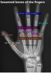
- Location and structure: Erica Chu, Donald Resnick. MRI Web Clinic — June 2014: Sesamoid Bones: Normal and Abnormal. Retrieved on 2017-11-04.
- Prevalences: Chen W, Cheng J, Sun R, Zhang Z, Zhu Y, Ipaktchi K et al. (2015). "Prevalence and variation of sesamoid bones in the hand: a multi-center radiographic study.". Int J Clin Exp Med 8 (7): 11721-6. PMID 26380010. PMC: 4565393.