Uploads by Boghog (usurped)
Jump to navigation
Jump to search
For Boghog (usurped) (talk · contributions · Move log · block log · uploads · Abuse filter log)
This special page shows all uploaded files that have not been deleted; for those see the upload log.
| Date | Name | Thumbnail | Size | Description |
|---|---|---|---|---|
| 18:22, 24 August 2008 | 1E5W.png (file) |  |
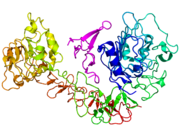
419 KB | {{Information |Description=Crystallographic structure of the N-terminal domain of moesin based on the {{PDB|1E52}} coordinates. |Source=self-made using PyMol |Date=2008-08-24 |Author= Boghog |Permission= |other_versions= }} [[Category |
| 19:56, 22 August 2008 | 2DDU.png (file) |  |
530 KB | {{Information |Description={{en|1=Crystallographic structure of the reelin protein based on the {{PDB|2DDU}} coordinates. Self made using PyMol.}} |Source=Own work by uploader |Author=Boghog |Date=2008-08-22 |Permission= |other_versions= } |
| 19:30, 22 August 2008 | 2e26.png (file) |  |
531 KB | Information |Description=Crystallographic structure of the reelin protein based on the {{PDB|2e26}} coordinates. |Source=self-made using PyMol |Date=2008-08-22 |Author= Boghog |Permission= |other_versions= }} Catego |
| 17:12, 16 August 2008 | 1NQL.png (file) |  |
635 KB | {{Information |Description={{en|1=Cartoon diagram of the epidermal growth factor receptor (EGFR) (rainbow colored, N-terminus = blue, C-terminus = red) complexed its ligand epidermal growth factor (magenta) based on the {{PDB|1NQL}} [[X-ra |
| 18:29, 12 August 2008 | 1H6F.png (file) |  |
558 KB | {{Information |Description=Crystallographic structure of the TBX3 protein dimer (cyan and green) complexed with DNA (brown) based on the {{PDB|1h6f}} coordinates. |Source=self-made using PyMol |Date=2008-08-12 |Author= [[User |
| 17:50, 9 August 2008 | CP154526.png (file) |  |
32 KB | {{Information |Description={{en|1=Chemical structure of CP154526, a Corticotropin-releasing hormone receptor antagonist.}} |Source=Own work by uploader. Created myself using ChemDraw. |Author=Boghog |Date=2008-08-09 |Permission= |other |
| 11:40, 9 August 2008 | 3CQV.png (file) |  |
701 KB | {{Information |Description=Cartoon diagram of the ligand binding domain of Rev-ErbA beta (rainbow colored, N-terminus = blue, C-terminus = red) complexed with heme (Space-filling model, carbon atoms = white, nitrogen = blue, oxygen = r |
| 07:17, 7 August 2008 | 2NEF.png (file) |  |
296 KB | {{Information |Description={{en|1=NMR structure of the NEF (protein based on the {{PDB|2NEF}} coordinates.}} |Source=Own work by uploader |Author=Boghog |Date=2008-08-07 |Permission= |other_versions= }} {{ImageUpload|full}} [[Category |
| 01:04, 6 August 2008 | 1BPV.png (file) |  |
284 KB | {{Information |Description={{en|1=crystallographic structure of the protein titin based on {{PDB|1BPV}}.}} |Source=Own work by uploader using PyMol |Author=Boghog |Date=2008-08-06 |Permission= |other_versions= }} {{ImageUpload|full}} |
| 00:01, 5 August 2008 | FOLATE PATHWAY1.png (file) |  |
42 KB | {{Information |Description=Folate metabolism pathway. |Source=self-made based on FOLATE_PATHWAY1.jpg |Date=2008-08-05 |Author= Boghog |Permission= |other_versions= }} Category:Biochemical cycles |
| 00:06, 3 August 2008 | 1AQD.png (file) |  |
429 KB | {{Information |Description={{en|1=Structure of HLA-DRA (cyan) complexed with HLA-DRB1 (green) and a fragment of HLA-A (magenta) based on the {{PDB|1AQD}} coordinates.}} |Source=Own work by uploader using PyMol |Author=Boghog |D |
| 23:26, 2 August 2008 | 2Q6W.png (file) |  |
421 KB | {{Information |Description={{en|1=Structure of HLA-DRA (cyan) complexed with HLA-DRB3 (green) and Integrin, beta 3 (magenta) based on {{PDB|2Q6W}} coordinates.}} |Source=Own work by uploader using PyMol |Author=Boghog |Date=2008-08-03 |Per |
| 23:00, 2 August 2008 | 1FV1.png (file) |  |
450 KB | {{Information |Description=Structure of HLA-DRA (cyan) complexed with HLA-DRB5 (green) and MBP (magenta) based on {{PDB|1FV1}} coordinates. |Source=self-made using pymol |Date=2008-08-03 |Author= Boghog |P |
| 10:25, 17 July 2008 | 1GCC.png (file) |  |
374 KB | {{Information |Description={{en|1=NMR structure of the GCC-BOX binding domain of EREBP (green) complexed with DNA (brown) based on {{PDB|1GCC}}.}} |Source=Own work by uploader created using PyMol based o |
| 15:12, 16 July 2008 | 1UVQ.png (file) |  |
464 KB | {{Information |Description=Crystallographic structure of HLA-DQA1 (cyan) complexed with HLA-DQB1 (green) and HCRT (magenta) based on {{PDB|1uvq}}. |Source=self-made |Date=2008-07-16 |Author= Boghog |Permission= |other_versions= |
| 19:45, 10 June 2008 | Estriol v2.png (file) |  |
49 KB | {{Information |Description={{en|1=3,16,17-estriol}} |Source=Own work by uploader |Author=Boghog |Date= |Permission= |other_versions= }} Created myself using ChemDraw. {{ImageUpload|full}} |