Neural development
Jump to navigation
Jump to search
Diagram
[edit]-
Human Cortical Development. Gyrification, gyration/sulcation, cortical folding, cortical convolution, fissuration or fissurization.
-
Slices through the brain at the mid-ventricular level and at the level of the centrum semiovale from six of the eight MR images obtained between 26 and 39 week gestational age
Timeline in human
[edit]3 weeks
[edit]-
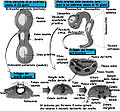
Human embryo between eighteen and twenty-one days old.
4 weeks
[edit]-
Brain of a four-week old human embryo.
-
Section of medulla spinalis of a four weeks’ embryo.
-
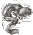
Exterior of brain of human embryo of four and a half weeks.
-
Brain of human embryo of four and a half weeks, showing interior of fore-brain.
-
Human embryo from thirty-one to thirty-four days.
5 weeks
[edit]-
Exterior of brain of human embryo of five weeks.
-
Interior of brain of human embryo of five weeks.
-
Human Embryo (7th week of pregnancy, 5th week p.o.)
6 weeks
[edit]-
Diagram of the various parts of the brain of a six-week old embryo.
-
A diagram showing the brain and major nerves of a 6 week old human fetus.
7 weeks
[edit]-
9-Week Human Embryo from Ectopic Pregnancy (7th week p.o.)
3 months
[edit]-
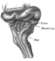
Hind-brain of a human embryo of three months—viewed from behind and partly from left side.
-
Median sagittal section of brain of human embryo of three months.
4 months
[edit]-
Inferior surface of brain of embryo at beginning of fourth month.
-
Median sagittal section of brain of human embryo of four months.
5 months
[edit]-
Outer surface of cerebral hemisphere of human embryo of about five months.


