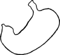
L'Estomac et le Corset
Jump to navigation
Jump to search
English: « L’Estomac et le Corset » is a French thesis about the damages from the corset on the stomach.
Français : « L’Estomac et le Corset » est un thèse français sur les dommages du corset sur l’estomac.
Images
[edit]-
Page 17:Français : Figure 1 (d’après Boas) ; THORAX ET ESTOMAC NORMAUX ; - Bord inférieur du foie. Diaphragme. G Gros intestin.English: Figure 1 (according to Boas) ; NORMAL THORAXES AND STOMACH ; - lower Edge of the liver Diaphragm. G Large intestine
-
Page 21 ;Français : Figure 2 (d’après Ziemssen) ; MOULE EN PLATRE D’UN ESTOMAC NORMAL.
-
Page 22 ;Français : Figure 3 (d’après Rosenheim) ; ESTOMAC NORMAL, INSUFFLÉ; - Bord inférieur du foie.
-
Page 42 ;Français : Figure 4 APPAREIL DE JAWORSKI ; pour l’insufflation de l’estomac
-
Page 49 ;Français : Figure 5 MONTRANT LA DÉFORMATION DU THORAX PAR LE CORSET; Fille de 27 ans ayant porté un corset serré. Estomac vertical. Capacité un peu augmentée. Pylore abaissé. - Bord inférieur du foie.
-
Page 54 ;Français : Figure 6 (d’après Dickinson); FORMES ET SITUATION NORMALES DU TRONC, DE L’ABDOMEN, DU FOIE ET DES ORGANES GÉNITO-URINAIRES. P. Poumon. - F. Foie.
-
Page 54 ;Français : Figure 7 (d’après Dickinson) DÉFORMATION PAR LE CORSET; Poitrine soulevée et reportée en haut. Ventre plus proéminent. Déformation du foie.
-
Page 55 ;Français : Figure 8 (d’après Dickinson); COUPE TRANSVERSALE DU THORAX AU NIVEAU DE L’ÉPIGASTRE
-
Page 58 ;Français : Figure 9 SCHÉMA MONTRANT LA DÉFORMATION D’UN ESTOMAC PINCÉ ENTRE UN FOIE ET UNE RATE HYPERTOPHIÉS; O. Ombilic.
-
Page 58 ;Français : Figure 10 d’après un moulage en plâtre de Ziemssen); ESTOMAC VERTICAL NON DILATÉ; mais présentant la même déformation, moins accusée, que celui de la fig. 9.
-
Page 62 ;Français : Figure 11 SCHÉMA REPRÉSENTANT LA MARCHE PROGRESSIVE DE LA DÉVIATION; O. Ombilic;
-
Page 62 ;Français : Figure 12 ESTOMAC VERTICAL CYLINDRIQUE (Moule en plâtre de Ziemssen).
-
Page 63 ;Français : Figure 13 ESTOMAC VERTICAL EN PLACE (Ziemssen). Commencement de poche sous-pylorique.
-
Page 64 ;Français : Figure 14 ESTOMAC VERTICAL CYLINDRIQUE; Dessiné d’après nature.
-
Page 65 ;Français : Figure 15 SCHÉMA DE LA DÉFORMATION DE LA POCHE SOUS-PYLORIQUE; Le pointillé indique la progression de l’ectasie. O. Ombilic;
-
Page 66;Français : Figure 16 (d’après nature). Le côté droit du thorax a été laissé schématisé pour montrer la déformation du thorax.English: Figure 16 (according to nature). The right side of the thorax was left schematized to show the deformation of the thorax.
-
Page 67;Français : Figure 17 ESTOMAC VERTICAL (2e degré). Demi-schéma d’après nature. D. Déformation thoracique.
-
Page 68;Français : Figure 18 (d’après nature). ESTOMAC VERTICAL AVEC BILOCULATION. D. Déformation thorax, correspondant au rétrécissement de l’estomac. On voit les côtes déformées, faisant un angle en avant de la ligne axillaire; ellers sont très-inclinées en bas.
-
Page 69;Français : Figure 19 (d’après Rosenheim). POSITION VERTICALE DE L’ESTOMAC, DE DIMENSIONS NORMALES. Sa situation pendant le gonflement, - Bord inférieur du foie.
-
Page 70;Français : Figure 20 (d’après Rosenheim). Dislocation totale d’un estomac normal. Gonflement artificiel. Le caria est abaissé. Vaste poche sous-pylorique due au mouvement de bascule de l’estomac en bas, et plutôt à droite qu’à gauche. - Bord inférieur du foie.
-
Page 71;Français : Figure 21 (d’après Rosenheim). ESTOMAC DILATÉ, INSUFFLÉ. Cette figure montre une vaste dilation sans dislocation; le pylore est presque à sa place. Elle présente un heureux point de comparaison avec les estomacs dilatés et disloqués en même temps.
-
Page 76;Français : Figure 22 (d’après Ziemssen). ECTASIE CONSIDÉRABLE. TENDANCE A LA VERTICALITÉ.
-
Page 82;Français : Figure 23


