Category:H&E stain
(Redirected from H&E stain)
histological stain method using hematoxylin and eosin | |||||
| Upload media | |||||
| Instance of |
| ||||
|---|---|---|---|---|---|
| Subclass of |
| ||||
| |||||
Hematoxylin & eosin stain.
Media in category "H&E stain"
The following 81 files are in this category, out of 81 total.
-
01064cha.tif 3,398 × 3,091; 30.52 MB
-
Auge Entwicklung.jpg 3,908 × 2,547; 2.41 MB
-
Batería de tinción hematoxilina eosina.jpg 873 × 623; 48 KB
-
Breast DCIS histopathology (1).jpg 500 × 376; 108 KB
-
Cartilage polarised.jpg 700 × 525; 68 KB
-
Cartilage.jpg 237 × 174; 9 KB
-
Cartilage01.JPG 4,000 × 3,000; 1.3 MB
-
Cartilage02.JPG 4,000 × 3,000; 1.44 MB
-
Cartilage03.JPG 4,000 × 3,000; 1.3 MB
-
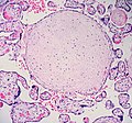
Cartilaginous metaplasia in placenta (?) 1 (590387907).jpg 1,470 × 1,375; 638 KB
-
Cartilaginous metaplasia in placenta (?) 2 (590739268).jpg 1,767 × 1,512; 969 KB
-
Cholestasis 2 high mag.jpg 2,848 × 4,272; 3.58 MB
-
Cholestasis 2 intermed mag.jpg 2,688 × 3,928; 3.4 MB
-
Cholestasis 2 low mag.jpg 2,448 × 4,272; 2.51 MB
-
Cholestasis high mag.jpg 4,272 × 2,848; 3.79 MB
-
Eosinophilic, basophilic, chromophobic and amphophilic staining.png 993 × 985; 1.05 MB
-
Figure 1a H&E staining in UC.tif 1,077 × 900; 2.43 MB
-
Figure 3a H&E staining in NSCLC.tif 1,080 × 900; 2.68 MB
-
Figure 4a H&E staining in NSCLC.tif 1,080 × 900; 2.74 MB
-
H&E Eby Loyola University Chicago.tif 794 × 794, 2 pages; 1.83 MB
-
Hair follicles.jpg 2,592 × 1,944; 1.87 MB
-
Histopathology of cytoplasmic hypereosinophilia in a pituitary adenoma.jpg 1,259 × 939; 389 KB
-
Immunohistochemistry technologies with AI.png 730 × 346; 419 KB
-
Kleinhirn Katze - 450x - HE-Färbung.jpg 640 × 480; 129 KB
-
Lagenidium giganteum f. caninum.jpg 4,096 × 3,000; 1.63 MB
-
Langerhanssche Insel.jpg 596 × 518; 126 KB
-
Micrographs (10.3897-zookeys.801.23088) Figure 6.jpg 1,512 × 1,786; 1.28 MB
-
Musc est long 400.JPG 4,000 × 3,000; 7.7 MB
-
Musc liso 400.JPG 4,000 × 3,000; 1.38 MB
-
Muscular estriado transv 400X.JPG 4,000 × 3,000; 2.49 MB
-
Muskel ( 1).jpg 3,886 × 2,578; 5.69 MB
-
Olfactory Epithelium Thin Section 1.5 microns H & E Stain Ward's 2.jpg 1,536 × 2,048; 668 KB
-
Olfactory Epithelium Thin Section 1.5 microns H & E Stain Ward's 4.jpg 1,536 × 2,048; 725 KB
-
Parasite140027-fig2 Histological sections of Dictyocoela diporeiae.tif 1,654 × 1,240; 3.97 MB
-
Parasite140076-fig1 Dirofilaria repens removed from a subcutaneous nodule - Photos.png 1,645 × 2,894; 5.31 MB
-
Parasite140085-fig3 Echinococcus granulosus cysts from boiled livers and lungs of sheep.tif 2,039 × 2,998; 4.78 MB
-
Parasite140105-fig1 Toxoplasmosis in a bar-shouldered dove - histology.tif 1,654 × 2,057; 3 MB
-
Parasite140105-fig2 Toxoplasmosis in a bar-shouldered dove - histology.tif 1,378 × 1,117; 828 KB
-
Parasite160015-fig5 - Small intestines of suckling mice inoculated or not.png 1,771 × 491; 1.63 MB
-
Parasite160019-fig3B-Chromidina spp. (Oligohymenophorea).png 950 × 1,349; 1.8 MB
-
Proctitis with reactive changes - alt -- high mag.jpg 2,848 × 4,272; 4.81 MB
-
Proctitis with reactive changes -- high mag.jpg 2,848 × 4,272; 4.94 MB
-
Proctitis with reactive changes -- intermed mag.jpg 2,848 × 4,272; 5.57 MB
-
Radiation proctitis - 2 -- high mag.jpg 2,848 × 4,272; 4.33 MB
-
Radiation proctitis - 2 -- intermed mag.jpg 4,272 × 2,848; 4.8 MB
-
Radiation proctitis - 2 alt -- high mag.jpg 4,272 × 2,848; 4.82 MB
-
Radiation proctitis - alt -- low mag.jpg 2,848 × 4,272; 5.01 MB
-
Radiation proctitis -- high mag.jpg 2,848 × 4,272; 5.03 MB
-
Radiation proctitis -- intermed mag.jpg 2,848 × 4,272; 5.57 MB
-
Radiation proctitis -- low mag.jpg 4,272 × 2,848; 5.46 MB
-
Radiation proctitis -- very high mag.jpg 2,848 × 4,272; 4.95 MB
-
Radiation proctitis -- very low mag.jpg 4,272 × 2,848; 4.31 MB
-
Rat hair follicles and vessels.jpg 2,592 × 1,944; 1.91 MB
-
Rat hair follicles.jpg 2,828 × 2,916; 2.76 MB
-
Rat skin (derma).jpg 2,592 × 1,944; 3.4 MB
-
Scalp cross section (negro).jpg 3,740 × 3,740; 7.89 MB
-
Scalp cross section (negro)2.jpg 3,557 × 3,557; 7.88 MB
-
Secretory phase endometrium.jpg 2,040 × 1,536; 1.08 MB
-
Setup for HE staining of frozen section slides.jpg 3,953 × 2,237; 2.55 MB
-
Trichinella rat BAM1.jpg 336 × 300; 72 KB
-
Urothelial carcinoma in situ - alt -- high mag.jpg 4,272 × 2,848; 4.28 MB
-
Urothelial carcinoma in situ -- high mag.jpg 4,272 × 2,848; 5 MB
-
Urothelial carcinoma in situ -- intermed mag.jpg 4,272 × 2,848; 5.81 MB
-
Urothelial carcinoma in situ -- very high mag.jpg 4,272 × 2,848; 4.54 MB
-
Whole slide image of H&E stained breast tumour tissue.png 8,213 × 5,321; 34.44 MB


