File:Viruses-12-00020-g004 PBCV-1 CryoEM (frameless).png

Original file (2,617 × 2,562 pixels, file size: 5.34 MB, MIME type: image/png)
Captions
Captions
Summary
[edit]| DescriptionViruses-12-00020-g004 PBCV-1 CryoEM (frameless).png |
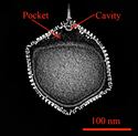
English: CryoEM structure of PBCV-1. (A) Hexagonal arrays of major capsomers form trisymmetrons and pentasymmetrons. The unique vertex with its spike structure is at the top. Capsomers in neighboring trisymmetrons are related by a 60° rotation, giving rise to the boundary between trisymmentrons. A fiber extents from one of the capsomers in each of the trisymmetrons. (B) Central cross-section of the cryo-EM density. A pocket is located between the virus internal membrane and the unique vertex and a cavity is located at the bottom of the spike structure. (C) PBCV-1 attached to the cell wall of its host chlorella as viewed by quick-freeze, deep etch microscopy. Note the virions are attached to the wall by fibers. (D) The cryoEM density (3.5 Å resolution) of PBCV-1 after removing the major capsid protein so that 14 minor proteins are visible. Each protein is shown in a different color as indicated on the right. |
| Date | |
| Source |
https://www.mdpi.com/viruses/viruses-12-00020/article_deploy/html/images/viruses-12-00020-g004.png at https://www.mdpi.com/1999-4915/12/1/20/htm (edit) Viruses 2020, 12(1), 20;doi:10.3390/v12010020 This article belongs to the Special Issue Viruses Ten-Year Anniversary. Licensee MDPI, Basel, Switzerland. This article is an open access article distributed under the terms and conditions of the Creative Commons Attribution (CC BY) license (https://creativecommons.org/licenses/by/4.0/). |
| Author | Provided by James L. Van Etten, Irina V. Agarkova, David D. Dunigan |
| Other versions |
 |
Licensing
[edit]- You are free:
- to share – to copy, distribute and transmit the work
- to remix – to adapt the work
- Under the following conditions:
- attribution – You must give appropriate credit, provide a link to the license, and indicate if changes were made. You may do so in any reasonable manner, but not in any way that suggests the licensor endorses you or your use.
- share alike – If you remix, transform, or build upon the material, you must distribute your contributions under the same or compatible license as the original.
File history
Click on a date/time to view the file as it appeared at that time.
| Date/Time | Thumbnail | Dimensions | User | Comment | |
|---|---|---|---|---|---|
| current | 19:26, 12 March 2021 |  | 2,617 × 2,562 (5.34 MB) | Ernsts (talk | contribs) | Uploaded a work by Provided by James L. Van Etten, Irina V. Agarkova, David D. Dunigan from https://www.mdpi.com/viruses/viruses-12-00020/article_deploy/html/images/viruses-12-00020-g004.png at https://www.mdpi.com/1999-4915/12/1/20/htm (edit) Viruses 2020, 12(1), 20;doi:10.3390/v12010020 This article belongs to the Special Issue Viruses Ten-Year Anniversary. Licensee MDPI, Basel, Switzerland. This article is an open access article distributed under the terms and conditions of the Creativ... |
You cannot overwrite this file.
File usage on Commons
The following 5 pages use this file:
Metadata
This file contains additional information such as Exif metadata which may have been added by the digital camera, scanner, or software program used to create or digitize it. If the file has been modified from its original state, some details such as the timestamp may not fully reflect those of the original file. The timestamp is only as accurate as the clock in the camera, and it may be completely wrong.
| Horizontal resolution | 236.2 dpc |
|---|---|
| Vertical resolution | 236.2 dpc |


