File:How-to-evaluate-the-microcirculation-report-of-a-round-table-conference-cc6118-S4.ogv
Jump to navigation
Jump to search
Size of this JPG preview of this OGG file: 800 × 450 pixels. Other resolutions: 320 × 180 pixels | 640 × 360 pixels | 1,024 × 576 pixels.
Original file (Ogg multiplexed audio/video file, Theora/Vorbis, length 12 s, 1,024 × 576 pixels, 1.96 Mbps overall, file size: 2.82 MB)
Captions
Captions
Add a one-line explanation of what this file represents
Summary
[edit]| Description |
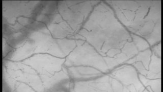
English: A video clip file showing severely altered microcirculation in a patient with sever sepsis. Forty-six small vessels (including 20 with absent flow and 14 with intermittent flow) and 15 large vessels (all perfused) are visualized. MFIs for each quadrant determined clockwise from the left upper one are 0, 0, 3 and 0. Accordingly, PPV is 15%, PVD is 1.4/mm and MFI is 0.75. |
||
| Date | |||
| Source | Video file from De Backer D, Hollenberg S, Boerma C, Goedhart P, Büchele G, Ospina-Tascon G, Dobbe I, Ince C (2007). "How to evaluate the microcirculation: report of a round table conference". Critical Care. DOI:10.1186/cc6118. PMID 17845716. PMC: 2556744. | ||
| Author | De Backer D, Hollenberg S, Boerma C, Goedhart P, Büchele G, Ospina-Tascon G, Dobbe I, Ince C | ||
| Permission (Reusing this file) |
This file is licensed under the Creative Commons Attribution 2.0 Generic license.
|
||
| Provenance |
|
File history
Click on a date/time to view the file as it appeared at that time.
| Date/Time | Thumbnail | Dimensions | User | Comment | |
|---|---|---|---|---|---|
| current | 10:45, 15 December 2014 | 12 s, 1,024 × 576 (2.82 MB) | Open Access Media Importer Bot (talk | contribs) | Automatically uploaded media file from Open Access source. Please report problems or suggestions here. |
You cannot overwrite this file.
File usage on Commons
There are no pages that use this file.