File:Evidence-of-Flicker-Induced-Functional-Hyperaemia-in-the-Smallest-Vessels-of-the-Human-Retinal-pone.0162621.s001.ogv
Jump to navigation
Jump to search
Size of this JPG preview of this OGG file: 800 × 422 pixels. Other resolutions: 320 × 169 pixels | 640 × 338 pixels | 1,024 × 540 pixels | 1,488 × 785 pixels.
Original file (Ogg Theora video file, length 17 s, 1,488 × 785 pixels, 524 kbps, file size: 1.04 MB)
Captions
Captions
Add a one-line explanation of what this file represents
Summary
[edit]| Description |
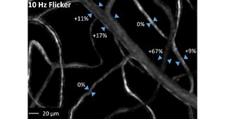
English: Animation of pre- and post-flicker images from the region of interest shown in Fig 2 . This region contains a venule and associated capillaries. Alternating between the registered pre- and post-flicker images aids visualization of vessel diameter changes induced by localized flicker stimulation. |
||
| Date | |||
| Source | S1 Video from Duan A, Bedggood P, Bui B, Metha A (2016). "Evidence of Flicker-Induced Functional Hyperaemia in the Smallest Vessels of the Human Retinal Blood Supply". PLOS ONE. DOI:10.1371/journal.pone.0162621. PMID 27617960. PMC: 5019460. | ||
| Author | Duan A, Bedggood P, Bui B, Metha A | ||
| Permission (Reusing this file) |
This file is licensed under the Creative Commons Attribution 4.0 International license.
|
||
| Provenance |
|
File history
Click on a date/time to view the file as it appeared at that time.
| Date/Time | Thumbnail | Dimensions | User | Comment | |
|---|---|---|---|---|---|
| current | 20:53, 12 October 2016 | 17 s, 1,488 × 785 (1.04 MB) | Open Access Media Importer Bot (talk | contribs) | Automatically uploaded media file from Open Access source. Please report problems or suggestions here. |
You cannot overwrite this file.
File usage on Commons
The following page uses this file:
Transcode status
Update transcode statusMetadata
Categories:
- Videos of cardiovascular anatomy
- Videos of blood vessels
- Blood flow
- Ocular system
- Videos of anatomy of the eye
- Videos of electron and ion microscopes and microprobes
- Scanning electron microscopy
- Retinal vessels
- Videos of cell biology
- Videos of cell types
- Videos of animal cells
- Videos of blood cells
- Videos of red blood cells
- Media from PLOS ONE