File:Aves gastrulation Origins of flow organisation.jpg

Original file (1,660 × 1,579 pixels, file size: 661 KB, MIME type: image/jpeg)
Captions
Captions
Summary
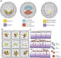
[edit]| DescriptionAves gastrulation Origins of flow organisation.jpg | Fig. 3. Origins of flow organisation. A) Flow organisers. From left to right: Attractors and repellers of the tissue flow, territories of directed and stochastic cell intercalation and myosin cables B) Cell behaviours that drive epithelial tissue flows, cell intercalation, cell division and ingressions. Division and ingression also contribute to cell rearrangements since they create changes of epithelial topology. C) Mechanism of contraction propagation based on tension dependent stabilisation of apical junctional myosin. From top to bottom: 1) The apical contraction of an individual cell due to a local accumulation of myosin, 2) results in the stretching of neighbouring cell junctions. The increase in tension produced by the stretching leads to the local stabilisation of myosin in these cells resulting in 3) apical contraction in these cells and propagation to further neighbours. |
| Date | |
| Source | https://www.sciencedirect.com/science/article/pii/S0925477320300290 Cellular processes driving gastrulation in the avian embryo |
| Author | Guillermo Serrano Nájera, Cornelis J. Weijer |

|
This file, which was originally posted to an external website, has not yet been reviewed by an administrator or reviewer to confirm that the above license is valid. See Category:License review needed for further instructions.
|
Licensing
[edit]- You are free:
- to share – to copy, distribute and transmit the work
- to remix – to adapt the work
- Under the following conditions:
- attribution – You must give appropriate credit, provide a link to the license, and indicate if changes were made. You may do so in any reasonable manner, but not in any way that suggests the licensor endorses you or your use.
Rights and permissions: https://s100.copyright.com/AppDispatchServlet?publisherName=ELS&contentID=S0925477320300290&orderBeanReset=true
Creative Commons
This is an open access article distributed under the terms of the Creative Commons CC-BY license, which permits unrestricted use, distribution, and reproduction in any medium, provided the original work is properly cited.
You are not required to obtain permission to reuse this article. [[Category:Aves]
File history
Click on a date/time to view the file as it appeared at that time.
| Date/Time | Thumbnail | Dimensions | User | Comment | |
|---|---|---|---|---|---|
| current | 16:55, 15 April 2024 |  | 1,660 × 1,579 (661 KB) | Rasbak (talk | contribs) | {{Information |description=Fig. 3. Origins of flow organisation. A) Flow organisers. From left to right: Attractors and repellers of the tissue flow, territories of directed and stochastic cell intercalation and myosin cables B) Cell behaviours that drive epithelial tissue flows, cell intercalation, cell division and ingressions. Division and ingression also contribute to cell rearrangements since they create changes of epithelial topology. C) Mechanism of contraction propagation based on ten... |
You cannot overwrite this file.
File usage on Commons
There are no pages that use this file.
Metadata
This file contains additional information such as Exif metadata which may have been added by the digital camera, scanner, or software program used to create or digitize it. If the file has been modified from its original state, some details such as the timestamp may not fully reflect those of the original file. The timestamp is only as accurate as the clock in the camera, and it may be completely wrong.
| JPEG file comment | HiRes |
|---|