File:41396 2020 879 Fig2.webp
From Wikimedia Commons, the free media repository
Jump to navigation
Jump to search

Size of this PNG preview of this WEBP file: 788 × 600 pixels. Other resolutions: 316 × 240 pixels | 631 × 480 pixels | 1,010 × 768 pixels | 1,280 × 974 pixels | 2,015 × 1,533 pixels.
Original file (2,015 × 1,533 pixels, file size: 657 KB, MIME type: image/webp)
File information
Structured data
Captions
Captions
Add a one-line explanation of what this file represents
Summary
[edit]| Description41396 2020 879 Fig2.webp |
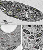
English: Visualization of intracellular bacteria Ca. Bodocaedibacter vickermanii (Holosporales) in Bodo saltans (Kinetoplastida). Images from Fluorescent in situ hybridisation (FISH) experiment (A–D), transmission electron microscopy (TEM) experiment (E–G) and Serial Block Face-Scanning Electron Microscopy (H, I). Imaging of B. saltans cell cultures with differential interference contrast (A), staining with DAPI to visualize nucleus and kinteoplast (B), FISH staining with endosymbiont specific probe conjugated to Cy3 (C) and images from three channels overlaid (D). Images were acquired on a Zeiss Axio Observer Z1 (Carl Zeiss AG, Jena, Germany) equipped with 100×1.4NA objective, 2.5x optovar. Images captured were analyzed using ImageJ v2.0. Ultrastructure of the B. saltans cell (E) displaying nucleus (nuc), kinetoplast (kp), mitochondria (mt), food vacuole (fv), flagellar pocket (fp) and three intracellular bacteria marked with dark red arrows, endosymbionts showing the presence of an electron lucid halo around cell membrane (F, G). 3D model of B. saltans cellular ultrastructure from two different angles (H, I). Bean shaped B. saltans cell is shown with flagellar pocket (in orange). Nucleus (in yellow) is situated in the centre of the cell and cytobionts (in blue) are distributed in close vicinity to the nucleus. A large, swirled mitochondrion (in magenta) is placed around the periphery of the cell, with a kinetoplast capsule just under the flagellar pocket. Multiple food vacuoles (in green) are present at the posterior end of the cell. |
||
| Date | |||
| Source | Fig. 2 at Bodo saltans (Kinetoplastida) is dependent on a novel Paracaedibacter-like endosymbiont that possesses multiple putative toxin-antitoxin systems. In: The ISME Journal 15, pages 1680–1694; doi:10.1038/s41396-020-00879-6 | ||
| Author | Samriti Midha, Daniel J. Rigden, Stefanos Siozios, Gregory D. D. Hurst, Andrew P. Jackson | ||
| Other versions |
|
Licensing
[edit]This file is licensed under the Creative Commons Attribution-Share Alike 4.0 International license.
- You are free:
- to share – to copy, distribute and transmit the work
- to remix – to adapt the work
- Under the following conditions:
- attribution – You must give appropriate credit, provide a link to the license, and indicate if changes were made. You may do so in any reasonable manner, but not in any way that suggests the licensor endorses you or your use.
- share alike – If you remix, transform, or build upon the material, you must distribute your contributions under the same or compatible license as the original.
File history
Click on a date/time to view the file as it appeared at that time.
| Date/Time | Thumbnail | Dimensions | User | Comment | |
|---|---|---|---|---|---|
| current | 06:49, 26 March 2023 |  | 2,015 × 1,533 (657 KB) | Ernsts (talk | contribs) | Uploaded a work by Samriti Midha, Daniel J. Rigden, Stefanos Siozios, Gregory D. D. Hurst, Andrew P. Jackson from Fig. 2 at ''''Bodo saltans'' (Kinetoplastida) is dependent on a novel ''Paracaedibacter''-like endosymbiont that possesses multiple putative toxin-antitoxin systems''. In: ''The ISME Journal'' 15, pages 1680–1694; doi:10.1038/s41396-020-00879-6 with UploadWizard |
You cannot overwrite this file.
File usage on Commons
The following 3 pages use this file:
Structured data
Items portrayed in this file
depicts
15 January 2021
image/webp
d7eab2477717606b5cdc3eead9450a3689d8c058
673,258 byte
1,533 pixel
2,015 pixel
Hidden categories:


