Commons:Wiki Science Competition 2023/Winners
Evaluation process
[edit]For more information about the selection procedure please go here.
National finalists
[edit]In this table you can find all information about national finalists (and eventually, national winners) of WSC2023.
Click on these page to know all information related to local jurors, timelines and finalists.
Argentina · Estonia · Finland · France and Monaco · Indonesia · Ireland · Italy · Malaysia · Nigeria · North Macedonia · Poland · Russia · South Africa · Spain · Switzerland · Ukraine · United States ··· The rest of the World
People in Science · Microscopy images · Non-photographic media · Wildlife & nature · Astronomy · General category · Image sets ··· Winners
Second international round
[edit]The national finalists are selected in an intermediate round focused mostly on the originality and the image quality.
- Explanation method
Similarly to Wiki Loves Monuments, international and national juries can show different taste, especially when some juries are small. For this reason it possible that some second finalists are given a higher ranking than the national winners at the international level.
Winners
[edit]The final winner and 4-6 runners-up were finally selected out of a final brainstorming from the finalists after the intermediate round, with some minor rearrangement based on
- the quality of their scientific description;
- the resolution and the presence of technical and scientific details;
- specific strong criticism by jurors or the academic committee (e.g. very common idea);
- use by wikimedia community (as of Ocotber 2024);
 Used on other wikimedia platforms[1]
 Featured file or
  Featured media
 Quality images
 Valued file
- excessive similarity to the winners and runners-up of the previous edition;
- a final balance of subjects and themes.
Please notice that in order to maximize the chance of every file, and due to specific taste of national and international juries, some files can be categorized twice.[2]
People in Science
[edit]
|
I am a lawyer based in Switzerland and photography is my favorite hobby. I've been lucky enough to travel a lot and that's what made me start photography, as it is such a great way of creating beautiful memories that can be seen again. In 2017 I got a taste for hiking and the magnificent natural landscapes that Mother Nature has to offer and that soon afterwards led me to take up landscape photography. After a hike during which I came into contact with wild ibexes, I enjoyed photographing them and very quickly developed a real passion for wildlife photography. Through my photographs I hope to share with the world the beauty of nature and animals. Some wild animals are very difficult to approach without disturbing them, and I often wear a ghillie suit when I go looking for them. By remaining undetected, I can observe animal behaviour that would otherwise not be observable if the animal would have had knowledge of human presence.The camouflage also allows me to get closer to animals and get good photographs without disturbing them. My main goal of the day when this picture was taken was to search for a group of wolves that was seen multiple times in the previous weeks in the area and that attacked a group of cows. Unfortunately I didn’t see wolves that day but that’s just nature! |
| International runner-up | International runner-up | |||
| Fritz Gorodiski (Fritz Goro), circa 1976. |
Children in a village of Purulia (West Bengal, India) playing and experimenting with a foldscope, an optical microscope that can be assembled with a sheet of paper and a lens. |
|||
| International runner-up | International runner-up | |||
| Covered snow pit in Antarctica illuminated by sun light shining through the snow from an adjacent pit. The chemicals and gases in the layers record the climate conditions. |
Sampling for microbiology from geothermally heated soils at Tramway Ridge, Mt. Erebus, Antarctica. |
|||
| International runner-up | International runner-up | |||
| Researcher checking the growth of oil palm seedlings placed in a controlled temperature environment. |
Drilling peat bog in Southern Estonia; the peak thickness can reach up to 10 meters but in this small bog in Karula national park the peat thicknesn is about 7 meters.. |
|||
Wildlife and Nature
[edit]
|
I am a software engineer based in Spain and photography is one of my main hobbies. I’ve been taking photos for most of my life, always fascinated by the ability to freeze a moment in time and being able to relive it later. I also use it to counteract my bad memory. I was not always a fan of showing my works, deeming them average. It took self-reflection, lots of trial and error and finally, pursuing a degree in photography and video, before I decided to start sharing them with my closest ones. Actually, it was my partner Estela who pushed me to submit some of them to this competition. The story behind this specific picture is one of cold weather, firewood and foggy mornings. At the time, we lived in a little house in the middle of the forest. Several times a day we would go out to the woodshed to collect logs for the fire that warmed our home. One day, we spotted a huge black and yellow salamander inside the shed. Not far from it, was this little clutch of eggs inside a small hole in the ground. I took my tripod and tried to get as close as possible to it. I didn’t have a macro lens at hand so I even tried inverting the lens while holding it, but the depth of field was too thin. So, I relied on my 18-35mm while using my phone flash and a hiking headlamp as my sources of light. After taking some pictures, we covered the hole partially with a log to avoid stepping on them. Day after day we visited to see the progress but sadly, we missed the day the eggs hatched. |
| International runner-up | International runner-up | |||
| A collection of crane flies hanging from their nest. |
Macro Shot of the face of a Giant peacock moth caterpillar. |
|||
| International runner-up | International runner-up | |||
| Coryphella sp., a nudibranch gastropod (Otranto, Lecce - Italy). |
Common Blue Damselflies (Enallagma cyathigerum) mating.. |
|||
| International runner-up | International runner-up | |||
| The close-up of a full color Galaxea spp in the Sulu Sea. |
Hover Fly (possibly Fazia (Allograpta) micrura) on a wild sage (Salvia verbenaca). |
|||
Microscopy images
[edit]
|
Since I was a little girl, I knew I wanted to understand why leaves are green, how fish swim and how the heart beats. One of the first childhood memories I have is the day my parents took me with five years old to visit the Museum of Natural History of Paris, and I saw this huge whale skeleton suspended in the air. I dreamed about it for years, telling myself that I had to understand the origin of life. Later, my passion for photography and traveling, made the perfect link with my work. My specialty is cell biology, and more specifically the study of basic cell mechanisms and embryo development using marine models. The ocean has always fascinated me, because its organisms have completely magical ways of adapting to extreme environments. On top of that, they can be useful for understanding fundamental biological mechanisms that also apply to humans! Currently working at the Institut Jacques Monod in the Nicolas Minc Team (CNRS, Paris), I'm using sea urchin embryos to study the mechanisms that regulate the orientation of cell divisions, a phenomenon linked to human cancer development. This is exactly what we see in this microscopy acquisition. In turquoise we see the membranes of dividing cells, in red the microtubules that serve as “ropes” to pull the chromosomes, and in green the DNA in the form of chromosomes. |
| International runner-up | International runner-up | |||
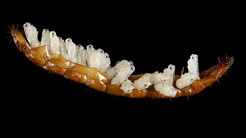
| Pupae of the Paracodrus apterogynus in the shell remains of a click beetle (Elateridae) larva. |
Image of five Toxoplasma gondii parasites (tachyzoites) in a human foreskin fibroblast, obtained using ultrastructure expansion microscopy. |
|||
| International runner-up | International runner-up | |||
| Mating of Boeckella gracilis copepods observed in dark field microscopy. |
Confocal image of a fruit fly retina expressing a toxic form of the RdgB protein, leading to degeneration. |
|||
| International runner-up | International runner-up | |||
| SEM Image of ZnO nanorod structures taken from UKM research laboratory. |
Differentiation of Human-Induced Pluripotent Stem Cells to GABAergic Neurons. |
|||
General category
[edit]
|
I am a professor from Ukraine, PhD, DrSci, specializing in mycology, evolutionary and molecular biology. I am the author of around 400 scientific publications, as well as popular books on the history of art. Photography has been a lifelong passion of mine, though I see it not as a separate art but rather as a way to share my impressions about the beauty of the world. As a professional, I often photograph my scientific subjects using macro photography, light, and even electron microscopy. For pleasure, I capture architecture, classical art, and living things around me. During a scientific expedition to Australia in March 2023, I was fortunate to find chains of glowing droplets in a deep niche of a rocky cliff. I already knew they were one of the wonders of Australian fauna, the traps of predatory fungus gnat larvae, and I tried to take a good photograph. However, the fact that this particular photo received such high recognition came as a huge surprise to me, since capturing it did not require significant effort. Nonetheless, these creatures are truly stunning!. |
| International runner-up | International runner-up | |||
| View of a human skull with roots in orbits and nasal cavities. |
Omocestus viridulus, known as the common green grasshopper. |
|||
| International runner-up | International runner-up | |||
| Icebreaker against the ice in Antarctica. |
An intense cloud-to-ground lightning strike near Goldendale, Washington. |
|||
| International runner-up | International runner-up | |||
| Hektoen enteric agar plate on which a stool sample has been cultured, exhibiting both lactose fermentation (orange) and hydrogen sulfide production (black). |
Mount Merapi's glowing lava slide. |
|||
Non-photographic media
[edit]
|
My name is Maxim Bilovitsky, I am a popularizer of science from Estonia, and also a blogger on YouTube. For one of my videos, I decided to show the transition of carbon dioxide to a supercritical state, for which I bought a small ampoule with this substance. In this ampoule, carbon dioxide is under high pressure, about 70 atmospheres, which is why it is in liquid form. If you fix such an ampoule and heat it with an ordinary hair dryer above 32 degrees Celsius, then the carbon dioxide in the ampoule will go into a supercritical state, which will cause the phase separation line in the ampoule to disappear. After some time, the ampoule begins to gradually cool down to room temperature, which is why the carbon dioxide inside again goes into a liquid state, but not immediately, since the walls of the ampoule have some heat capacity. From this, you can observe such an unusual effect of changing the phase state of the substance.
|
| International runner-up | International runner-up | |||
| Video of the volcano eruption next to Litli-Hrútur in Iceland in 2023. |
Synthetic images of the Moon using orthographic projections. |
|||
| International runner-up | International runner-up | |||
| Sublimation of iodine under a microscope. |
Melting an iron nail with a supercapacitor. |
|||
| International runner-up | International runner-up | |||
| Video of silver micromirrors in solution under optical darkfield microscope demonstrating Brownian motion, Casimir effect and colorful scattering of surface plasmons. |
Starlink satellites passing over the Swiss night sky as seen from Mürren. |
|||
Astronomy
[edit]
|
Since as far back as I can remember, I've been interested in the stars, and some of my earliest memories are of looking up at the skies and asking my elders "why?" as kids tend to do. This is what led to me being a scientist. Since age 7, when I got my first telescope, I've been an amateur astronomer. In 1986, when Halley's Comet was visible, I was 13 and I learned to build my own reflecting telescope at that time, but technology wasn't good enough for the average person to capture images of what I saw. I was not a rich student in college so my hobby took a back seat, but once I began my professional career, I started using my disposable income to buy a lot of telescopes and technology finally caught up to enable amateurs to produce amazing images. All this took off in high gear when we moved to rural western New York on Lake Ontario in 2014, which has fairly dark skies. It was a few years of getting progressively better: I was a national finalist in 2019, but later on I was able to produce some of my best images ever, which I submitted to the 2021 Wiki Science Competition where I was named an international winner. However, following that productive period, between travel during the summer and weather not cooperating, my imaging had slowed down. I also focussed more on quality instead of quantity and with better processing so there were fewer submissions this time around to the 2023 Wiki Science Competition. The Lion Nebula is an extended emission nebula in the constellation Cepheus. Its apparent distance is more than 10,000 light years away from Earth. While it fits nicely within the FOV of my 4" scope reduced to 490mm focal length, it is estimated to be about 250 light years across. (For reference, the diametre of our planet Earth is 0.0425 light seconds across!) The Lion Nebula (particularly the parts outside the "head") is very faint, which is why I spent so much time on this target, and I was trying hard to get the mesh-like body below the head and outer structures and filaments which shows up the most with the O3 filter on which I collected ~24 hours worth of data. Most of the images shown were processed by first creating an original LHOO/SHO image, removing the stars, light processing, and then putting the stars back at a reduced size. Overall, this was a challenging image to process but the effort is reasonably worth it. Professionally, I am currently a computational biology and bioinformatics Professor at the University at Buffalo researching multiscale modelling of macromolecular structure, function, interaction, design, and evolution at multiple scales.. |
| International runner-up | International runner-up | |||
| Horsehead Nebula IC434 and Flame Nebula NGC2024. |
Flying Bat and Squid nebulae Sh2-129/Ou4 narrowfield (m-sho, c-shorgb/131h). |
|||
| International runner-up | International runner-up | |||
| Astrophotography of the NGC 3324 nebula in narrowband technique and Hubble palette (SHO). |
Astrophotography of the NGC6188 nebula in narrowband technique and Hubble palette (SHO). |
|||
| International runner-up | International runner-up | |||
| A cosmic odyssey captured over 525 hours of dedicated amateur astrophotography efforts, unveiling the majesty of Cassiopeia A's supernova remnant. |
Astrophotography of M20 nebula in LRGB technique. |
|||
Image sets
[edit]
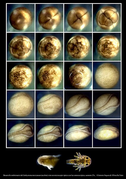
In many Wiki Science Competition editions, microscopy plays a crucial role in the "Image Sets" category by revealing details hidden to the naked eye. A series of images tells a more complete and compelling story. This image series showcases the developmental journey of an axolotl embryo, from its earliest stages to a fully formed organism. The precision and clarity of the images highlight not only the beauty and complexity of developmental biology but also the meticulous techniques involved in sample preparation and imaging. The symmetry and thoughtful layout enhance the clarity and impact of the series, adding an element of visual elegance. |
|
My wife and I, both co-authors of the photography, are undergraduate students in Biological Sciences at the National University of Comahue in Bariloche, Argentina. We began delving into the microscopic world during the pandemic, as we could not attend labs in person. To continue our studies, we acquired a microscope to conduct lab work at home. Having access to the microscope allowed us to dedicate many hours to microscopic observation and improving photography techniques. Currently, we have had the opportunity to learn and use a wide variety of techniques and microscopes, such as confocal microscopy, scanning electron microscopy, and refining techniques like polarized light, dark field, and phase contrast microscopy. Additionally, we have designed 3D-printed parts to adapt various photography devices to conventional microscopes, enabling us to capture high-quality images that contribute to the scientific dissemination of the microscopic world. The story behind the photograph is quite interesting. As biology students, we are deeply drawn to peculiar animals, which led us to the opportunity to raise a pair of axolotls (Ambystoma mexicanum). Although axolotls are critically endangered in the wild, there are more individuals in captivity worldwide than in their natural habitat, which in some way helps preserve the species. After a year of care, our axolotls reproduced, giving us the chance to document the embryonic development of an axolotl egg. To achieve this, the axolotl egg remained under the microscope for 18 days with constant hydration around the clock. Being so delicate, it could not be moved from one place to another, so it stayed under the microscope throughout the entire development process. |
| International runner-up | International runner-up | |||
| Organisms found in water. |
Microbial Collection. Cell culture plates researched in foods. |
|||
| International runner-up | International runner-up | |||
| Argulus foliaceus. |
||||
| International runner-up | International runner-up | |||
. |
Analysis of collagen expression in young Salamanders (P. waltl) via HCR FISH visualization. |
|||
Notes
[edit]- ↑ in main namespaces, ns0, that is content-related pages
- ↑ For example some users might prefer the "wildlife and nature" category, but some landscapes are also considered under "general", similarly certain jury accept images in the "people in science" category where only a portion of the body is visible, so they are sometimes included in other categories as well (es: general) at the international level.
- ↑ Special Prize WMCH for the "Best third position"
- ↑ a b c Special Prize WMCH for "Climate Sciences"


