Commons:Wiki Science Competition 2019/Winners/Microscopy images/round 2
Jump to navigation
Jump to search
Selection from the national finalists.
 Featured file
 Quality images
 Valued file
 Used on wikimedia platforms
currently voting: 10 out of 12.
See also round 1
-
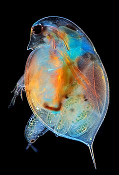
Cladoceran from Daphnia genus giving birth.
 MarekMiś
10.9
7.708 out of 10, SD 1.982

-
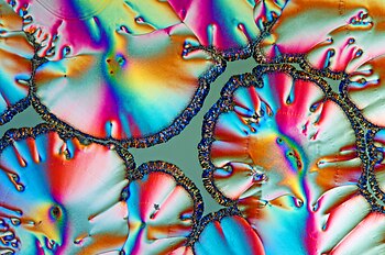
Crystals, mainly sugar, in a dried Coca Cola droplet, taken with a microscope using cross polarization
 Alexander Klepnev
11.1
7.083 out of 10, SD 2.984

-
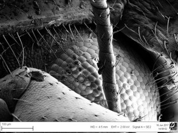
eye of Chrysopidae (stacking of 30 frames)
 Грибков михаил
11.2
6.042 out of 10, SD 2.911

-
"Coffee ring" from Aluminium Oxide particles (150 nm) on cover glass coated with gold (thickness 20 nm)  Мохаммед Аль-Музайкер, Таир Есенбаев
13.3
5.208 out of 10, SD 3.447 -
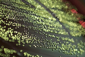
Escherichia coli growing on Eosin methylene blue (EMB) media with its distinctive green metallic sheen
 Gene Drendel
13.4
5.833 out of 10, SD 2.219 -
Spontaneous mutation occured during regeneration of Schmidtea mediterranea.
 Marcinekenator
13.9
6.042 out of 10, SD 2.911
-
Surface oxides of conductive 3D copper structures are visible under the microscope as a blueish glimmer.
 Davidpervan
14.0
6.875 out of 10, SD 3.220 -
Larva of Culex Pipiensìì seen with polarizing microscopic technique
 Massimo Brizzi
14.3
7.083 out of 10, SD 2.575 -
Mouse cortical neurons
 ALol88
14.3
6.667 out of 10, SD 2.683
-
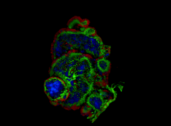
3D projection of a Patiria miniata bipinnaria
 Natalie Carrigan
14.7
7.083 out of 10, SD 2.087
-
TRIAP1 regulats P53 expression in HTC116 mitochodrial compartiments.
(int.) GAbderrahmane
15.0
5.417 out of 10, SD 2.345 -
Parasitic rider's head (size 1 mm)
 Грибков михаил
15.0
6.458 out of 10, SD 3.100 -
Tongue of a common house fly, flaring out
 C1bill
15.1
6.667 out of 10, SD 2.462 -
Characteristic growth pattern of Bacillus mycoides growing on agar supplemented with food dye colouring.
 Gene Drendel
15.2
5.000 out of 10, SD 2.611 -
Sphalerite from the Irish base metal ore field.
 Aileen Doran
16.1
5.417 out of 10, SD 2.344 -
Microscopic view of salt crystals from a dried human tear.
 Arseniy1109
16.3
5.833 out of 10, SD 2.683 -
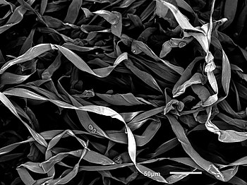
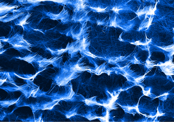
Fibers lining the inside of an acorn shell detected using a scanning electron microscope (SEM)
 Marissa Dessellier
16.4
6.250 out of 10, SD 2.500 -
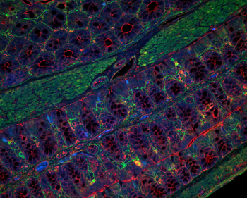
Immunofluorescence staining showing proximal part of the large intestine from 8 weeks old mouse.
 Abhimanu Pandey
16.5
7.500 out of 10, SD 2.132 -
Block nanostructures on the surface of the p-InP after photoelectrochemical etching in the electrolyte 12H2O+2HCl+1HBr
 Suchikova Yana & Kovachov Sergey
16.9
5.208 out of 10, SD 3.278 -
Scanning Electron Microscopy (SEM) image showing TiO2 nanowires produced by hydrothermal synthesis.
 M.T. Buscaglia
17.2
6.250 out of 10, SD 2.2613 -
Tartaric acid crystals under polarized light
 MarekMiś
17.3
6.667 out of 10, SD 3.257 -
SEM-image of the textured indium phosphide surface.
 Яна Сычикова
17.5
6.250 out of 10, SD 2.919 -
Butterfly wing under 20x magnification.
 Tanel Nook
17.8
6.459 out of 10, SD 2.251 -
Transmission electron microscope image of a tightly-focused laser beam, showing the individual crests and troughs of its electromagnetic wave.
 J. J. Axelrod
19.5
5.625 out of 10, SD 3.0386 -
Cytoskeleton of human fibroblast (red, F-actin stained with TRITC Phalloidin) after internalization of polydopamine nanoparticles (Green, lipophilic dye)
 Matteo Battaglini
19.9
5.833 out of 10, SD 1.628 -
Ladybug's eye
 Léonito 2003
20.0
6.250 out of 10, SD 3.286
-
Ascorbic acid crystals under polarized light
 MarekMiś
20.0
6.875 out of 10, SD 2.845
-
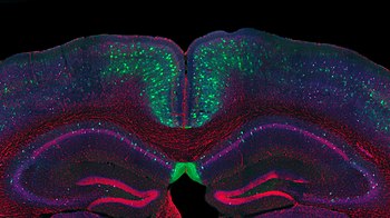
A coronal view of mouse hyppocampus reveals the nuclei density (in red) and the extracellular matrix structures that envelope neurons (in green).
 Luisa Demarchi, Ilaria Bertocchi, Alessandra Oberto
21.2
6.250 out of 10, SD 2.718 -
Hydroxyapatite overgrowing the biomaterial.
 Jan Ozimek
21.2
5.0 out of 10, SD 2.820 -
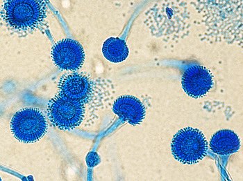
Aspergillus fumigatus
 Azienda Ospedaliera SS. Antonio e Biagio e Cesare Arrigo, Alessandria
21.8
5.000 out of 10, SD 2.132 -
Scanning Electron Microscope (SEM) view of a natural sponge
 Micropoz
22.3
5.000 out of 10, SD 3.844 -
Immunofluorescence image of intestinal organoids generated using small intestine from ApcMin/+ mice, which carry an Apc mutation leading to the spontaneous development of intestinal tumours
 Abhimanu.pandey
22.4
5.833 out of 10, SD x -
10 𝝻m heart-shaped sample under scanning election microscopy (SEM).
 Yating Zhang
22.4
5.208 out of 10, SD 3.100 -
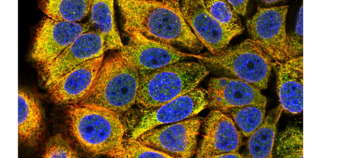
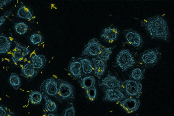
Pathogenic bacterium Pseudomonas aeruginosa (yellow) infects and kills a cell culture of lung epithelial cells (blue).
 Benoit-Joseph Laventie
23.5
6.667 out of 10, SD 2.219
-
Scanning electron micrograph of the dorsal side of a newly hatched Eisenia hortensis.
 Hannah G Watson, Andrew T Ashchi, Glen S Marrs, Cecil J Saunders
23.5
5.625 out of 10, SD 3.555
-
Colorized (pseudo-colors) electron microscopy (SEM) image of lilac blossom
 Smirnov Evgeny
28.0
6.875 out of 10, SD 2.165
Comments
[edit]- This year with tried to discriminate using also the ranking phase, not only the average with standard deviation.
