Commons:Wiki Science Competition 2017/Winners/Estonia
Jump to navigation
Jump to search
Algeria · Australia · Austria · Bangladesh · Belgium · Brazil · Bulgaria · Chile · China · Colombia · Czech Republic · Egypt · Estonia · France · Georgia · Germany · Greece · India · Indonesia · Iran · Iraq · Ireland · Italy · Jordan · Mexico · Oman · Philippines · Poland · Romania · Russia · Saudi Arabia · Serbia · Singapore · Spain · Sweden · Switzerland · Thailand · Ukraine · United Arab Emirates · United Kingdom · United States ··· The rest of the World
![]() These are the finalists for WSC2017 in Estonia.
These are the finalists for WSC2017 in Estonia.
This country-level selection had a national organizer (Ivo Kruusamägi) and a national coordination page.
Jury
[edit]- 144 files were submitted and all of them were considered valid
- The finalists were selected on 2017-12-22
- The jury was composed of Jaak Kikas, Ivo Kruusamägi, Ulvar Käärt, Arko Olesk, and Urmas Tartes
- The results were published on 2018-02-11
Finalists
[edit]People in science
[edit]| Category winner | National finalist | |||
| Assistant professor Ivar Arold in the summer of 1990 during field work with geography students of University of Tartu, explaining levelling. Margus Hendrikson |
Characterizing snow and ice in snow pits at Fox Fonna glacier, Svalbard. Based on snow density, grain size and structure as well as snow hardness, it is possible to characterize the weather conditions that have occurred before in the location. Kertu Liis Krigul |
| ||
| National finalist | National finalist | |||
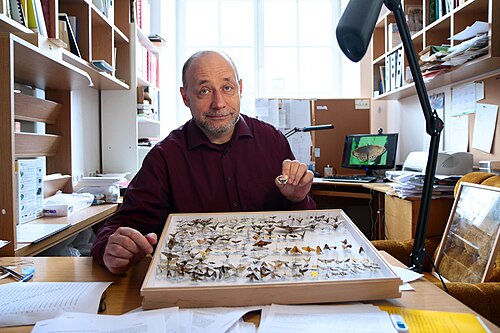
| Photo of an entomologist, professor Toomas Tammaru of University of Tartu, in his office. Lauri Kulpsoo |
Photo of the professor of entomology taking down a light trap used to catch butterflies. Donald McPart |
| ||
| National finalist | ||||
| Ester Oras removing a tooth from a mummy in the Art Museum of the University of Tartu. The authenticity of the mummy was confirmed with radiocarbon dating and DNA profiling. Ester Oras & Mait Metspalu |
||||
Microscopy images
[edit]| Category winner | National finalist | |||
| “Nanocigarette”. Upon heating silica coated silver nanowires (diameter ~ 100 nm) to 600 °C silver (the brighter material on the images) starts to swell out of the silica shell and by doing so, starts to break the shell and accumulate at the end of the wire (creating a “cloud of smoke”). The thickness of the silica shell is only about 30 nm, so it is almost transparent for electrons and therefore it can be seen that some of the silver is still encapsulated in the shell. Mikk Vahtrus |
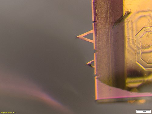
The cantilever of an atom force microscope (AFM) is used for force measurements between different substances. The photo is made with a stereo microscope just before the start of the experiment with AFM to see if both tips are still there, as they have a tendency to break off very easily. Kertu Liis Krigul |
|||
| National finalist | National finalist | |||
| Tongue under optical microscope. Striated muscle tissue - vertical- and cross-section. Olga Botsarova |
Crystals of yttrium barium copper oxide under a scanning electron microscope. Artur Kaljo, Karl Rene Kõlvart, Mari Kass |
| ||
| National finalist | ||||
| Polydicyanamide film near rubber sealing. Optical microscopy image, picture width 200 micrometers, brightfield contrast mode. It was prepared by electropolymerizing of dicyanamide-based ionic liquid during the testing of high voltage graphene capacitor technology. Tavo Romann |
||||
Non-photographic media
[edit]| Category winner | ||||
| Freezing of a water droplet at temperature of approximately -180 °C. Maxim Bilovitskiy |
||||
Image sets
[edit]| Category winner | National finalist | |||
| While aiming to create divergent white-light speckle pattern using a diffuser and a lens, the authors noticed a colorful wavy pattern on the wall. Patterns took various stunning forms while distance between the diffuser and the lens was changed. Heli Lukner, Sandhra-Mirella Valdma, Andreas Valdmann
|
Structure inside autoclaved aerated concrete (AAC) with various crystals (tobermorite, CaSO4, Jennite etc). Tarmo Tamm, Markus Otsus, Artur Kaljo |
|||
General category
[edit]| Category winner | National finalist | |||
| Two fluorescent dyes are injected into stationary upward water flow inside a three-dimensional porous medium made of a random stack of solid transparent beads of diameter 16 mm (the planar cross-sections of the spherical beads are seen as black circles on the image). Based on this single photograph, the evolution of the dye concentrations and their interaction rules in the porous medium have been deciphered and explained. Mihkel Kree |
The first finding of the southern bushcricket species Bicolorana bicolor in Estonia was in 2015. This is one of many signs that climate is changing. Photo from 2017. Veljo Runnel |
| ||
| National finalist | National finalist | |||
| The incubation chambers in Sassendalen, Svalbard. These chambers are used to indirectly assess Cyanobacteria's capability to fix N2 by enzyme nitrogenase. Kertu Liis Krigul |
Old karst landscape in Rõstla quarry (Central Estonia), exposed shortly during dolomite mining. It was formed before Ice-Age, and covered with till. Tõnu Pani |
| ||
| National finalist | ||||
| Waterspider Argyroneta aquatica and freshwater crustacean Asellus aquaticus at Rahaaugu nature protection area in Estonia. Lennart Lennuk |
||||


