Category:Veterinary histopathology
Jump to navigation
Jump to search
NOTE: Files should be categorized by species!
Subcategories
This category has the following 3 subcategories, out of 3 total.
Media in category "Veterinary histopathology"
The following 47 files are in this category, out of 47 total.
-
African swine fever infected macrophage.jpg 640 × 432; 66 KB
-
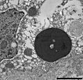
Apoptosis mouseliver.jpg 417 × 417; 49 KB
-
Apoptosis multi mouseliver.jpg 417 × 417; 43 KB
-
Apoptosis stained.jpg 485 × 477; 47 KB
-
Avian Pox TEM 01.tif 1,822 × 1,901; 2.72 MB
-
BIBD blood 121104-2-crop-v1.jpg 574 × 545; 139 KB
-
BIBD liver 092403-1 v1.jpg 791 × 596; 300 KB
-
BIBD TEM 0082916g004 v01.jpg 815 × 789; 360 KB
-
Birdflu2.jpg 600 × 502; 161 KB
-
Breast hyperplasia.jpg 960 × 717; 74 KB
-
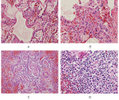
Breast of rat with carcinogen ana alkaloids.jpg 960 × 717; 52 KB
-
Breast of rat with carcinogen.jpg 960 × 717; 58 KB
-
Canine distemper pathology.jpg 500 × 643; 68 KB
-
Canine transmissible venereal tumor cytology.JPG 1,553 × 1,079; 194 KB
-
Chang IBD pancreas 0082916g005 v01.png 1,000 × 758; 1.9 MB
-
Chicken wire calcification chondroblastoma.jpg 1,280 × 720; 147 KB
-
Chytridiomycosis2.jpg 568 × 516; 47 KB
-
Dog skin cytology.jpg 436 × 423; 129 KB
-
Eastern equine encephalitis.jpg 700 × 876; 227 KB
-
Equine Protozoal Myeloencephalitis.jpg 329 × 336; 151 KB
-
Feline leukemia virus.JPG 1,808 × 1,200; 423 KB
-
Feline sporotrichosis 4.jpg 300 × 233; 14 KB
-
FIPCytology2.jpg 350 × 234; 40 KB
-
FIPHisto1.jpg 350 × 246; 66 KB
-
Giardia intestinalis dog.jpg 1,147 × 1,408; 148 KB
-
Grey horse melanoma 1.JPG 2,048 × 1,536; 3.24 MB
-
Jaagsiekte.jpg 1,200 × 1,801; 1.05 MB
-
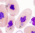
Leucocytozoon smithi.jpg 404 × 280; 20 KB
-
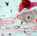
Malassezia tape cytology.jpg 370 × 279; 20 KB
-
Meyers b2 s0316 b3.png 187 × 292; 36 KB
-
Parasite140027-fig2 Histological sections of Dictyocoela diporeiae.tif 1,654 × 1,240; 3.97 MB
-
Pathology in CWD-Infected Animals.jpg 699 × 456; 58 KB
-
Phocine distemper encephalitis.jpg 600 × 388; 54 KB
-
Phocine distemper virus.jpg 600 × 382; 36 KB
-
Prototheca zopfii.jpg 2,163 × 1,449; 593 KB
-
Purkinje cell necrosis aviary encephalomyelitis.jpg 1,020 × 768; 92 KB
-
Rabies encephalitis Negri bodies PHIL 3377 lores.jpg 1,801 × 1,197; 438 KB
-
Rabies encephalitis PHIL 3368 lores.jpg 1,801 × 1,199; 635 KB
-
Rabies encephalitis PHIL 3368.png 1,801 × 1,199; 4.06 MB
-
Rabies negri bodies brain.jpg 700 × 453; 118 KB
-
Rabies Virus EM PHIL 1876.JPG 1,835 × 2,392; 2.33 MB
-
Sarcocystis in sheep oesophagus.JPG 2,304 × 1,536; 386 KB
-
Sarcocystis in sheep oesophagus2.JPG 1,911 × 1,536; 1.61 MB
-
Sarcoptic-mites-in-skin.jpg 1,883 × 1,939; 972 KB
-
Whirling disease pathology.jpg 1,099 × 736; 485 KB


