Category:Ultrasound
Jump to navigation
Jump to search
sound waves with frequencies above the human hearing range | |||||
| Upload media | |||||
| Audio | |||||
|---|---|---|---|---|---|
| Subclass of |
| ||||
| |||||
Subcategories
This category has the following 25 subcategories, out of 25 total.
A
B
- Bat echolocation (42 F)
D
- Dog whistles (12 F)
I
L
- Luxpole (4 F)
M
R
S
- Scanning acoustic microscopy (24 F)
- Ultrasonic stream gauges (1 F)
- Sylvatest (9 F)
T
U
- Ultrasonic cleaners (24 F)
- Ultrasonic flaw detection (39 F)
- Ultrasonic flaw detector (10 F)
- Ultrasonic flow meters (20 F)
- Ultrasonic therapy (4 F)
- Ultrasonography (42 F)
V
Media in category "Ultrasound"
The following 200 files are in this category, out of 269 total.
(previous page) (next page)-
1 Amplitude.jpg 270 × 570; 81 KB
-
1 focusing.gif 270 × 296; 220 KB
-
1 Source geometry.jpg 270 × 296; 20 KB
-
1-2 Стимуляция.jpg 529 × 884; 63 KB
-
10 Week Ultrasound 6.webm 9.1 s, 480 × 352; 1.12 MB
-
10 Week Ultrasound 7.webm 25 s, 480 × 352; 3.47 MB
-
15000Hz hugh pitch ultrasound.oga 30 s; 80 KB
-
18 Week Ultrasound 2.webm 44 s, 1,280 × 720; 13.83 MB
-
18 Week Ultrasound 3.webm 23 s, 1,280 × 720; 10.51 MB
-
18 Week Ultrasound 4.webm 1 min 7 s, 1,280 × 720; 29.9 MB
-
18 Week Ultrasound.webm 11 s, 1,280 × 720; 9.03 MB
-
2 Source geometry better.jpg 669 × 741; 65 KB
-
2-1г Набор излучателей.jpg 1,105 × 626; 81 KB
-
A medical ultrasound linear array probe, scan head, transducer.jpg 4,032 × 3,024; 4.12 MB
-
A-mode scan.png 300 × 200; 16 KB
-
Abdominal aorta ultrasound.jpg 716 × 397; 120 KB
-
Abdominal Ultrasound Full Exam 01.jpg 716 × 537; 126 KB
-
Abdominal Ultrasound Full Exam 02.jpg 716 × 537; 136 KB
-
Abdominal Ultrasound Full Exam 03.jpg 716 × 537; 136 KB
-
Abdominal Ultrasound Full Exam 04.jpg 716 × 537; 132 KB
-
Abdominal Ultrasound Full Exam 05.jpg 716 × 537; 140 KB
-
Abdominal Ultrasound Full Exam 06.jpg 716 × 537; 148 KB
-
Abdominal Ultrasound Full Exam 07.jpg 716 × 537; 141 KB
-
Abdominal Ultrasound Full Exam 08.jpg 716 × 537; 145 KB
-
Abdominal Ultrasound Full Exam 09.jpg 716 × 537; 141 KB
-
Abdominal Ultrasound Full Exam 10.jpg 716 × 537; 146 KB
-
Abdominal Ultrasound Full Exam 11.jpg 716 × 537; 142 KB
-
Abdominal Ultrasound Full Exam 12.jpg 716 × 537; 152 KB
-
Abdominal Ultrasound Full Exam 13.jpg 716 × 537; 147 KB
-
Abdominal Ultrasound Full Exam 14.jpg 716 × 537; 136 KB
-
Abdominal Ultrasound Full Exam 15.jpg 716 × 537; 154 KB
-
Abdominal Ultrasound Full Exam 16.jpg 716 × 537; 140 KB
-
Abdominal Ultrasound Full Exam 17.jpg 716 × 537; 140 KB
-
Abdominal Ultrasound Full Exam 18.jpg 716 × 537; 146 KB
-
Abdominal Ultrasound Full Exam 19.jpg 716 × 537; 123 KB
-
Abdominal Ultrasound Full Exam 20.jpg 716 × 537; 143 KB
-
Abdominal Ultrasound Full Exam 21.jpg 716 × 537; 127 KB
-
Abdominal Ultrasound Full Exam 22.jpg 716 × 537; 123 KB
-
Abdominal Ultrasound Full Exam 23.jpg 716 × 537; 135 KB
-
Abdominal Ultrasound Full Exam 24.jpg 716 × 537; 148 KB
-
Abdominal Ultrasound Full Exam 25.jpg 716 × 537; 139 KB
-
Abdominal Ultrasound Full Exam 26.jpg 716 × 537; 149 KB
-
Abdominal Ultrasound Full Exam 27.jpg 716 × 537; 162 KB
-
Abdominal Ultrasound Full Exam 28.jpg 716 × 537; 151 KB
-
Abdominal Ultrasound Full Exam 29.jpg 716 × 537; 151 KB
-
Abdominal Ultrasound Full Exam 30.jpg 716 × 537; 146 KB
-
Abdominal Ultrasound Full Exam 31.jpg 716 × 537; 138 KB
-
Abdominal Ultrasound Full Exam 32.jpg 716 × 537; 147 KB
-
Abdominal Ultrasound Full Exam 33.jpg 716 × 537; 140 KB
-
Abdominal Ultrasound Full Exam 34.jpg 716 × 537; 146 KB
-
Abdominal Ultrasound Full Exam 35.jpg 716 × 537; 151 KB
-
Abdominal Ultrasound Full Exam 36.jpg 716 × 537; 133 KB
-
Abdominal Ultrasound Full Exam 37.jpg 716 × 537; 148 KB
-
Abdominal Ultrasound Full Exam 38.jpg 716 × 537; 145 KB
-
Abdominal Ultrasound Full Exam 39.jpg 716 × 537; 163 KB
-
Abdominal Ultrasound Full Exam 40.jpg 716 × 537; 142 KB
-
Abdominal Ultrasound Full Exam 41.jpg 716 × 537; 138 KB
-
Abdominal Ultrasound Full Exam 42.jpg 716 × 537; 148 KB
-
Abdominal Ultrasound Full Exam 43.jpg 716 × 537; 214 KB
-
Abdominal Ultrasound Full Exam 44.jpg 716 × 537; 205 KB
-
Abdominal Ultrasound Full Exam 45.jpg 716 × 537; 136 KB
-
Abdominal Ultrasound Full Exam 46.jpg 716 × 537; 121 KB
-
Abdominal Ultrasound Full Exam 47.jpg 716 × 537; 134 KB
-
Abdominal Ultrasound Full Exam 48.jpg 716 × 537; 131 KB
-
Abdominal Ultrasound Full Exam 49.jpg 716 × 537; 133 KB
-
Abdominal Ultrasound Full Exam 50.jpg 716 × 537; 135 KB
-
Abdominal Ultrasound Full Exam 51.jpg 716 × 537; 132 KB
-
Abdominal Ultrasound Full Exam 52.jpg 716 × 537; 128 KB
-
Abdominal Ultrasound Full Exam 53.jpg 716 × 537; 122 KB
-
Abdominal Ultrasound Full Exam 54.jpg 716 × 537; 130 KB
-
Abdominal Ultrasound Full Exam 55.jpg 716 × 537; 137 KB
-
Abdominal Ultrasound General Illustration.jpg 1,552 × 970; 423 KB
-
Acoustically Levitated Objects in a TinyLev.jpg 700 × 378; 56 KB
-
AMG6-48228.png 1,604 × 1,174; 930 KB
-
Angle seam tofd.svg 800 × 600; 15 KB
-
Anterior compartment of left proximal leg.jpg 1,024 × 768; 157 KB
-
Araysonic6.jpg 1,350 × 600; 94 KB
-
Arduino Smart Stick.svg 881 × 569; 480 KB
-
ATL Philips HDI 3000 - HDI 5000 (15957734226).jpg 638 × 473; 62 KB
-
Barbell horn amplitude and stress distributions.jpg 423 × 206; 39 KB
-
Basketball autoscore with Arduino.svg 783 × 536; 580 KB
-
Bat bug eco.svg 793 × 517; 52 KB
-
Beam Function 1MHz.png 2,968 × 791; 55 KB
-
Befahrung Wismut-Stollen und Tiefer Elbstolln 2015-09-17 16.JPG 3,648 × 2,736; 2 MB
-
Chili 1.jpg 1,500 × 2,000; 787 KB
-
Chirps151018-21NR.mp3 1 min 5 s; 663 KB
-
Chirps151018-22NR.mp3 50 s; 491 KB
-
Chirps151018-23NR.mp3 23 s; 229 KB
-
Chirps190918-22s.mp3 22 s; 253 KB
-
Chirps190918-22s2.png 1,816 × 234; 750 KB
-
Chirps190918.mp3 50 s; 577 KB
-
Chirps20160512-50s.mp3 51 s; 594 KB
-
Chirps20160512.mp3 1 min 0 s; 722 KB
-
Clinical neuroimaging using ultrasound.svg 512 × 250; 692 KB
-
ColourFlowSonographicImagingSystemBlockDiagram-de.svg 728 × 339; 40 KB
-
Common Pipistrelle Echos 2020-04-21 (dkrb).ogg 14 s; 227 KB
-
Control box, courtesy of Ultrasonic Marine.jpg 1,803 × 1,682; 814 KB
-
Conventional Converging Horn.jpg 1,920 × 1,120; 275 KB
-
Converging horn amplitude and stress distributions.jpg 265 × 183; 27 KB
-
Coronal anterior section through anterior fontanelle using ultrasound.jpg 4,186 × 2,794; 3.3 MB
-
Curved Array Ultrasound Sensor Construction.jpg 5,257 × 3,263; 2.84 MB
-
DB Ultraschall-Schienenprüfzug 719-001 - 0 (Schild).JPG 2,144 × 2,343; 1.98 MB
-
Diagnostic Sylvatest.jpg 3,672 × 4,896; 5.48 MB
-
Diagram showing liver lesioning using a HIFU transducer 2.png 591 × 489; 50 KB
-
Diagram showing liver lesioning using a HIFU transducer.png 920 × 720; 209 KB
-
Dibujo Prinzip Ultraschall.PNG 442 × 422; 15 KB
-
Dibujo Ultraschall Gerinne.PNG 674 × 628; 28 KB
-
Dibujo Ultraschall2.PNG 690 × 597; 50 KB
-
Digitales-Ultraschallgerät.jpg 1,454 × 1,188; 1.11 MB
-
Doctor performs Ultherapy procedure.jpg 4,928 × 3,280; 1.12 MB
-
Dog ultrasound whistle ID tag.jpg 800 × 600; 63 KB
-
Dual element transducer.png 1,300 × 612; 70 KB
-
Echographe médical.jpg 4,032 × 3,024; 1.35 MB
-
Echographe Siemens à la clinique du bon secours Maroua.jpg 3,116 × 3,841; 1.49 MB
-
Efect piezoelectric.png 460 × 311; 8 KB
-
Ekkopuls.png 1,742 × 623; 85 KB
-
Emergency-department-ultrasonography-guided-long-axis-antecubital-intravenous-cannulation-How-to-do-2036-7902-4-3-S1.ogv 1 min 3 s, 640 × 480; 9.15 MB
-
-
-
Emetteur et 2 recepteurs ultrasons + mousse.jpg 2,560 × 1,440; 947 KB
-
Emetteur et 2 recepteurs ultrasons.jpg 2,560 × 1,440; 940 KB
-
Epiploic appendages 0001.jpg 717 × 486; 77 KB
-
Epiploic appendages 0002.jpg 637 × 414; 56 KB
-
Fetal Ultrasound numbers.png 1,200 × 1,111; 1.46 MB
-
Fetal Ultrasound.png 1,200 × 1,111; 1.28 MB
-
Fig3angleinsensitivity.gif 300 × 341; 13 KB
-
Fracture.jpg 574 × 433; 29 KB
-
Full-wave Barbell Horn.jpg 1,200 × 1,200; 244 KB
-
Gallbladder and common bile duct ultrasound.jpg 716 × 391; 127 KB
-
Garcia-Atance number.png 525 × 399; 17 KB
-
Gout signs ultrasound.jpg 472 × 410; 46 KB
-
Grading by ultrasound.png 1,058 × 642; 378 KB
-
Grafavgur.png 600 × 300; 7 KB
-
Gunaraj Awasthi3.jpg 650 × 400; 69 KB
-
Hepatofugal flow in portal vein.jpg 825 × 614; 45 KB
-
HighlandPICT RedJacket.jpg 4,000 × 3,000; 4.6 MB
-
Historische Ultraschallapparatur von Franziska Seidl.jpg 3,456 × 5,184; 7.64 MB
-
Horn transitional section shapes.jpg 500 × 278; 48 KB
-
HOROWITZ classification pericard effusion.jpg 1,754 × 2,480; 270 KB
-
How Ultrasound Imaging Works (Sonography).webm 1 min 41 s, 1,920 × 1,080; 8.89 MB
-
Hyperthermia Treatment For Cancer, Sonotherm 1000 by Labthermics.JPG 1,690 × 1,092; 587 KB
-
Il pipistrello come fa.ogg 3 min 9 s; 4.7 MB
-
Imaging phantom as seen on medical ultrasound.jpg 4,608 × 3,456; 5.22 MB
-
Installed ultrasonic transducer.tif 625 × 483; 392 KB
-
Linear striations of adenomyosis.jpg 554 × 412; 60 KB
-
Main applications and features of functional ultrasound (fUS) imaging.svg 512 × 439; 2.86 MB
-
Main brain functional imaging technique resolutions.svg 512 × 293; 320 KB
-
MASINT-UTAMStower.png 600 × 809; 465 KB
-
MASINT-WWI-SoundRanging.png 1,373 × 837; 47 KB
-
Maxim-1-HydrophonPZTinGehäuse.JPG 1,600 × 1,200; 768 KB
-
Mid saggital section of ultrasound cranium.jpg 4,608 × 3,456; 5.06 MB
-
Modeconversion.svg 499 × 431; 57 KB
-
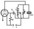
Montaj generator piezoelectric in rezonanta.png 317 × 268; 6 KB
-
Mounted OpenBikeSensor in Ulm, Germany.jpg 2,517 × 1,651; 995 KB
-
Networked ultrasonic sensors.jpg 615 × 327; 22 KB
-
Neuenrade - Niederheide - Gemeinschaftshauptschule 05 ies.jpg 3,888 × 2,592; 1.38 MB
-
Neuenrade - Niederheide - Gemeinschaftshauptschule 06 ies.jpg 3,888 × 2,592; 1.52 MB
-
Nonlinear US wave propagation.svg 512 × 244; 84 KB
-
Obstetric Ultrasound Polaroid Photograph 1985.jpg 2,522 × 2,010; 1.65 MB
-
OpenBikeSensor Montage am Gepäckträger.jpg 2,815 × 2,541; 2.08 MB
-
Opstelling ultrasoon onderzoek.PNG 480 × 198; 5 KB
-
Pancreas ultrasound doppler.jpg 716 × 499; 150 KB
-
Pancreas ultrasound.jpg 716 × 497; 131 KB
-
PhasArray1.jpg 470 × 954; 66 KB
-
Phased array beam.jpg 460 × 197; 31 KB
-
Phased array weld.jpg 455 × 330; 32 KB
-
Preclinical applications of fUS imaging.svg 512 × 307; 912 KB
-
Proloterapia guiada por ultrasonido.png 583 × 580; 28 KB
-
Proximity Meter with Sound Speed Calibration (schema).svg 497 × 559; 594 KB
-
Proximity Meter with Sound Speed Calibration.jpg 4,000 × 3,000; 3.68 MB
-
Proxxon distance meter.jpg 1,511 × 848; 328 KB
-
Pulse-echo method weld Seam.svg 800 × 600; 28 KB
-
REMS Raw unfiltered signals.png 1,281 × 678; 522 KB
-
REMS Segnali grezzi non filtrati.png 1,617 × 869; 945 KB
-
Renal cyst ultrasound 2.jpg 1,552 × 970; 388 KB
-
Renal cyst ultrasound 3.jpg 1,552 × 970; 211 KB
-
Renal cyst ultrasound.jpg 1,552 × 970; 417 KB
-
Resultado de medição com um analisador de frequência de transdutores.png 851 × 203; 151 KB
-
Reverb.svg 800 × 600; 28 KB
-
Rhythmodynamics Non-jet Propulsion.ogv 1 min 40 s, 1,280 × 720; 20.49 MB
-
Right kidney seen on abdominal ultrasound.jpg 1,552 × 904; 323 KB
-
Schéma décrivant le fonctionnement d’un bac à ultrasons.png 858 × 368; 56 KB
-
Sensore Parcheggio.png 851 × 560; 81 KB
-
Single-element source.jpg 217 × 206; 18 KB
-
Something you wouldn't see in America (cropped).jpg 1,732 × 1,075; 314 KB
-
Something you wouldn't see in America.jpg 2,080 × 1,544; 690 KB
-
Sound & Ultrasound frequencies.svg 887 × 374; 171 KB
-
Sound and ultrasound.jpg 5,683 × 2,500; 1.43 MB
-
Soundfield Water 4MHz TransducerRadius5mm.png 3,847 × 897; 68 KB
-
Southern Partnership Station 2016 Medical Team 160902-A-CP070-0305.jpg 2,432 × 1,824; 908 KB
-
Spleen ultrasound.jpg 716 × 501; 123 KB
-
ST - Chart 1.png 2,599 × 1,429; 108 KB
-
ST - Equation 1.png 2,065 × 821; 51 KB
-
ST - Equation 2.png 3,596 × 1,967; 201 KB


