Category:Taken with Olympus DP71
Jump to navigation
Jump to search
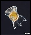
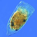
English: Taken with Olympus DP71, a camera designed to capture images from microscopes.
Wikimedia category | |||||
| Upload media | |||||
| Instance of | |||||
|---|---|---|---|---|---|
| Category combines topics | |||||
| photographs by photographic equipment used | |||||
photographs which have been taken by a specific camera | |||||
| Instance of | |||||
| |||||
Media in category "Taken with Olympus DP71"
The following 88 files are in this category, out of 88 total.
-
Alternaria alternata-5 copie.jpg 680 × 512; 124 KB
-
Capillaria hepatica adult 200x HB.jpg 300 × 300; 83 KB
-
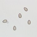
Capillaria hepatica eggs 200x HB.jpg 300 × 300; 82 KB
-
Carcinoma with enteroblastic differentiatin.jpg 1,360 × 1,024; 1.2 MB
-
Carcinoma with enteroblastic differentiation2-1.jpg 1,360 × 1,024; 1.18 MB
-
Carcinoma with lymphoid stroma.jpg 1,360 × 1,024; 1.41 MB
-
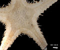
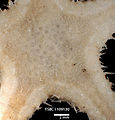
Cheiraster (Barbadosaster) echinulatus (13981096501).jpg 1,240 × 1,024; 206 KB
-
Cheiraster (Barbadosaster) echinulatus (13981107101).jpg 1,234 × 1,024; 187 KB
-
Cheiraster (Barbadosaster) echinulatus (13984285255).jpg 1,360 × 1,024; 167 KB
-
Cheiraster (Barbadosaster) echinulatus (13984288325).jpg 1,360 × 974; 190 KB
-
Cheiraster (Barbadosaster) echinulatus (13984617875).jpg 735 × 765; 120 KB
-
Chronic hypersensitivity pneumonitis - histology.jpg 1,360 × 1,024; 896 KB
-
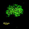
Cluster of PAOs.jpg 4,080 × 3,072; 3.18 MB
-
Comedo DCIS.jpg 1,360 × 1,024; 816 KB
-
DAPI Stained Biomass For phosphorus acclimating organism identification.jpg 4,080 × 3,072; 5 MB
-
Dinoflagellates and a tintinnid ciliate.jpg 3,878 × 2,870; 11.68 MB
-
Epiploctlis acuminata.jpg 569 × 700; 369 KB
-
Euphrosine triloba (13980884782).jpg 1,318 × 1,208; 186 KB
-
Euphrosine triloba (13980886262).jpg 1,153 × 1,424; 187 KB
-
Euphrosine triloba (13980906691).jpg 775 × 858; 114 KB
-
Fibroblast focus.jpg 2,040 × 1,536; 2.08 MB
-
Germ cell aplasia with focal maturation arrest.jpg 1,360 × 1,024; 692 KB
-
Gloeotrichia in Sytox.jpg 1,360 × 1,024; 450 KB
-
Granuloma 20x.jpg 1,360 × 1,024; 741 KB
-
Granuloma mac.jpg 1,360 × 1,024; 773 KB
-
Gyrodinium dinoflagellate.jpg 1,602 × 1,605; 2.91 MB
-
H&E 10x germ cell aplasia with focal maturation arrest.jpg 1,360 × 1,024; 941 KB
-
H&E 10x Invasive ductal.jpg 1,360 × 1,024; 867 KB
-
H&E 10x papilloma.jpg 1,360 × 1,024; 985 KB
-
H&E 20x hypospermatogenesis with peritubular fibrosis.jpg 1,360 × 1,024; 876 KB
-
H&E 4x hypospermatogenesis with peritubular fibrosis.jpg 1,360 × 1,024; 793 KB
-
Harmothoe sp (13984092915).jpg 1,136 × 706; 117 KB
-
Harmothoe sp (13984527274).jpg 1,272 × 1,329; 165 KB
-
Harmothoe sp (13984529214).jpg 1,011 × 613; 87 KB
-
Histology of chronic hypersensitivity pneumonitis.jpg 4,080 × 3,072; 3.75 MB
-
Honeycomb change.jpg 2,040 × 1,536; 1.91 MB
-
Hypolobated small megakaryocyte.jpg 1,360 × 1,024; 461 KB
-
Invasive Ductal Carcinoma 40x.jpg 1,360 × 1,024; 727 KB
-
Kurloff body, Guinea pig.jpg 1,822 × 1,463; 717 KB
-
Lineate Dovesnail (11407513074).jpg 391 × 846; 72 KB
-
Lineate Dovesnail (11671168853).jpg 382 × 855; 71 KB
-
Livoneca redmanii (13961082526).jpg 1,116 × 1,024; 193 KB
-
Livoneca redmanii (close-up of larvae in the brood pouch) (13961011896).jpg 1,360 × 1,024; 155 KB
-
Lobular carcinoma in situ.jpg 1,360 × 1,024; 735 KB
-
Lyngbya stained.jpg 804 × 654; 185 KB
-
Mecsina Etki Mekanizması .jpg 1,360 × 1,024; 457 KB
-
Melanoma 03.jpg 4,080 × 3,072; 4.58 MB
-
Microcystis in Sytox.jpg 648 × 648; 89 KB
-
Moderately differentiated tubular adenocarcinoma(tub2).jpg 1,360 × 1,024; 588 KB
-
Moniliformis moniliformis egg.jpg 300 × 300; 46 KB
-
Moniliformis moniliformis eggs.jpg 300 × 300; 25 KB
-
Necrogran10x.jpg 1,360 × 1,024; 1.01 MB
-
Oldest freshwater drum sagittal otolith.jpg 1,360 × 1,024; 780 KB
-
Ornithocercus heteroporus (probably).jpg 2,024 × 2,279; 2.25 MB
-
Ornithocercus Magnificus image.jpg 1,921 × 2,234; 2.74 MB
-
P. falciparum thick smear with gametocytes.jpg 1,360 × 1,024; 1.02 MB
-
P. falciparum thick smear with ring forms.jpg 1,360 × 1,024; 1,017 KB
-
P. falciparum thin smear gametocyte.jpg 1,360 × 1,024; 826 KB
-
P. falciparum thin smear ring forms and gametocyte.jpg 1,360 × 1,024; 833 KB
-
P. falciparum thin smear with ring forms.jpg 1,360 × 1,024; 846 KB
-
Pawsonaster parvus (13981509962).jpg 1,360 × 949; 168 KB
-
Pawsonaster parvus (13984709485).jpg 1,360 × 1,024; 190 KB
-
Pc12may28-MAY-28-2011.19.26.39-4.jpg 2,040 × 1,536; 1.22 MB
-
Poorly differentiated adenocarcinoma(por2).jpg 1,360 × 1,024; 693 KB
-
Poraniella echinulata (13985169234).jpg 1,260 × 1,024; 216 KB
-
Poraniella echinulata (13985173524).jpg 942 × 954; 170 KB
-
Poraniella echinulata.jpg 956 × 932; 167 KB
-
Processa profunda (11355307514).jpg 2,538 × 1,362; 236 KB
-
Roughback Shrimp (11356733713).jpg 1,139 × 676; 122 KB
-
S10-5263 H&E 20x DCIS.jpg 1,360 × 1,024; 934 KB
-
Snapping shrimp (13980940591).jpg 680 × 790; 86 KB
-
Snapping shrimp (13984113675).jpg 1,329 × 742; 120 KB
-
Snapping shrimp (13984556614).jpg 1,360 × 667; 100 KB
-
Spotted Slippershell (11358235156).jpg 1,215 × 897; 151 KB
-
Spotted Slippershell (11670983356).jpg 1,244 × 882; 142 KB
-
Star stick diatom.jpg 4,100 × 3,092; 4.88 MB
-
Striped Porcelain Crab (11355112454).jpg 2,613 × 2,384; 418 KB
-
Striped Porcelain Crab (11971406766).jpg 2,580 × 2,219; 435 KB
-
Synovial Sarcoma Biphasic High Power.jpg 1,360 × 1,024; 1.02 MB
-
Tethyaster grandis (13952984327).jpg 1,087 × 731; 123 KB
-
Tethyaster grandis (13961638746).jpg 917 × 946; 172 KB
-
Tethyaster vestitus (13961783946).jpg 713 × 1,024; 184 KB
-
Tethyaster vestitus (14004892993).jpg 1,041 × 674; 202 KB
-
Tintinnid ciliate Favella.jpg 2,400 × 2,400; 7.28 MB
-
UIPlungbiopsy.jpg 2,040 × 1,536; 1.83 MB
-
Winged Chimney Clam (11405003726).jpg 981 × 480; 85 KB
-
Winged Chimney Clam (11670624063).jpg 981 × 531; 79 KB
-
Winged Chimney Clam (11670754754).jpg 973 × 464; 79 KB


