Category:Spectroscopy
Jump to navigation
Jump to search
- (en) Spectroscopy
- (ar) مطيافية
- (bg) Спектроскопия
- (bs) Spektroskopija
- (ca) Espectroscòpia
- (cs) Spektroskopie
- (da) Spektroskopi
- (de) Spektroskopie
- (el) Φασματοσκοπία
- (eo) Spektroskopio
- (es) Espectroscopia
- (et) Spektroskoopia
- (fa) طیفسنجی
- (fi) Spektroskopia
- (fr) Spectroscopie
- (gl) Espectroscopia
- (he) ספקטרוסקופיה
- (hr) Spektroskopija
- (hu) Színképelemzés
- (id) Spektroskopi
- (it) Spettroscopia
- (ja) 分光法
- (ko) 분광학
- (lmo) Spetruscupia
- (ms) Spektroskopi
- (nl) Spectroscopie
- (nn) Spektroskopi
- (no) Spektroskopi
- (pl) Spektroskopia
- (pt) Espectroscopia
- (ro) Spectroscopie
- (ru) Спектроскопия
- (sh) Spektroskopija
- (simple) Spectroscopy
- (sk) Spektroskopia
- (sl) Spektroskopija
- (sr) Спектроскопија
- (su) Spéktroskopi
- (sv) Spektroskopi
- (ta) நிறமாலையியல்
- (th) สเปกโตรสโกปี
- (tr) Spektroskopi
- (ug) سپېكتروسكوپىيە
- (uk) Спектроскопія
- (ur) طیف بینی
- (vi) Phổ học
- (zh) 光谱学
measurement and interpretation of interactions between matter and electromagnetic radiation of varying frequencies | |||||
| Upload media | |||||
| Instance of |
| ||||
|---|---|---|---|---|---|
| Subclass of | |||||
| Different from | |||||
| |||||
Subcategories
This category has the following 48 subcategories, out of 48 total.
+
A
C
- Cauchy-Lorentz distributions (53 F)
D
- Dielectric spectroscopy (6 F)
- Dynamic light scattering (17 F)
E
F
- Fluorometers (27 F)
- Franck-Condon principle (23 F)
G
- Grotrian diagrams (9 F)
I
- Inverse photoemission (9 F)
M
- Muon spin spectroscopy (6 F)
N
- Nanoscale spectroscopy (7 F)
O
- Orgel diagrams (8 F)
P
S
- Spectroscopes (48 F)
- Spontaneous emission (24 F)
T
- Tanabe-Sugano diagrams (24 F)
V
X
Media in category "Spectroscopy"
The following 200 files are in this category, out of 434 total.
(previous page) (next page)-
12-NewStripRot90.jpg 5,000 × 1,000; 621 KB
-
13C std.svg 195 × 118; 30 KB
-
1911 Britannica-Argon-A. Schuster.png 439 × 142; 48 KB
-
2DCorrelationDemoDataset.svg 512 × 333; 43 KB
-
2DCorrelationSpectrum.svg 512 × 1,138; 31 KB
-
2DCorrelationSpectrumAC.svg 512 × 387; 18 KB
-
2DCorrelationSpectrumPresence.svg 512 × 512; 20 KB
-
2DIR pulse sequence.svg 512 × 259; 515 KB
-
3 D PICture.jpg 1,280 × 720; 53 KB
-
3- d picture final.jpg 606 × 394; 32 KB
-
3-D pic.png 859 × 533; 92 KB
-
4most scheme.jpg 2,510 × 1,500; 914 KB
-
Absorbance d'une solution en fonction du temps.JPG 653 × 349; 17 KB
-
Absorption or emission spectroscopy.png 600 × 400; 20 KB
-
Acerca de la espectroscopía (01H0FV2JD7QHG3BZRA42R2RE8K).jpg 4,000 × 6,600; 1.92 MB
-
Acerca de la espectroscopía (01H0FV2JD7QHG3BZRA42R2RE8K).png 4,000 × 6,600; 2.74 MB
-
Acerca de la espectroscopía (01H0FV2JD7QHG3BZRA42R2RE8K).tiff 4,000 × 6,600; 5.44 MB
-
AchtergrondSubtractie.png 577 × 268; 7 KB
-
Acoustic resonance spectroscopy diagram.png 588 × 626; 30 KB
-
Adip.PNG 835 × 520; 28 KB
-
Ald näide.png 419 × 327; 14 KB
-
Alpha300 Raman microscope.jpg 5,184 × 3,456; 4.81 MB
-
AmBe NRC ISO.jpg 3,299 × 1,952; 293 KB
-
Analyseur.png 1,010 × 673; 48 KB
-
Angular deviation MS.png 377 × 277; 11 KB
-
App3.jpg 3,120 × 4,160; 1.95 MB
-
Apsorpcija shema.png 1,356 × 294; 3 KB
-
Aqueous Potassium Ferricyanide Magnetic Circular Dichroism.svg 442 × 405; 187 KB
-
ASP3 003.jpg 2,992 × 2,992; 1.59 MB
-
AtomicLineAb.png 583 × 437; 9 KB
-
Base line no base line.jpg 1,453 × 890; 49 KB
-
Basic principle of Multi-Object Spectroscopy rearranged.png 714 × 760; 401 KB
-
Basic principle of Multi-Object Spectroscopy-ru.png 1,428 × 379; 451 KB
-
Basic principle of Multi-Object Spectroscopy.png 1,428 × 379; 644 KB
-
Bild Streakkamera.JPG 1,024 × 1,024; 153 KB
-
Bildschirmfoto 2016-09-19 um 10.30.41.png 1,320 × 968; 67 KB
-
Boltzmann distribution.png 400 × 300; 8 KB
-
BSA linearity.jpg 577 × 394; 30 KB
-
BSA Standard Curve.png 550 × 325; 23 KB
-
Bunsen spectrometer (Newth 1902).jpg 3,965 × 3,360; 3.05 MB
-
BZ Poissoning.png 2,164 × 2,108; 305 KB
-
Capron Rainband Spectrum.png 197 × 341; 84 KB
-
Cavity assisted spectroscopy principle.png 929 × 415; 37 KB
-
Cavity decay process.png 867 × 498; 27 KB
-
CF3I spectrum2.png 1,051 × 680; 34 KB
-
Characterization bench at LAAS 0479.jpg 3,872 × 2,592; 3.28 MB
-
Chemical ID with nano-FTIR.png 606 × 250; 50 KB
-
Chromophore.svg 921 × 532; 28 KB
-
CircularMultipassCell 17reflections.png 1,056 × 708; 125 KB
-
Cl-scheme.svg 641 × 425; 26 KB
-
Co2vibrations.gif 497 × 214; 9 KB
-
Color wheel wavelengths.png 740 × 550; 50 KB
-
Colourimeter.jpg 1,504 × 2,256; 2.42 MB
-
Combination plot CO.png 350 × 276; 2 KB
-
Complex Organic Molecules of NGC 1333 IRAS 2A Protostar (MIRI) (2024-111).png 3,840 × 2,766; 1.26 MB
-
Complex Organic Molecules of NGC 1333 IRAS 2A Protostar (MIRI) (2024-111).tiff 16,000 × 11,523; 44.46 MB
-
Confocal Raman microscope.jpg 2,275 × 2,657; 1.03 MB
-
Copper spectrum.png 383 × 75; 27 KB
-
Ct s1.png 900 × 735; 49 KB
-
Cubic elastic constants..JPG 383 × 158; 18 KB
-
Curve decomposition.svg 1,102 × 679; 58 KB
-
DART Measurements Plan.svg 1,052 × 744; 668 KB
-
Datacube MUSE on NGC 4650A with IFU.jpg 700 × 438; 33 KB
-
Derivative sum Lorentzians.png 427 × 313; 3 KB
-
Diag jab poziom.pdf 1,752 × 1,239; 63 KB
-
Diagram jablonskiego.svg 510 × 270; 14 KB
-
Diagramme états électroniques.png 2,048 × 830; 42 KB
-
Diatomic rigid rotor.svg 400 × 200; 25 KB
-
Dielectric responses pl.svg 454 × 370; 13 KB
-
Dielectric responses zh hans.svg 450 × 400; 47 KB
-
Dielectric responses zh hant.svg 450 × 400; 48 KB
-
Dielectric responses-ru.svg 737 × 638; 4 KB
-
Dielectric responses.svg 450 × 400; 35 KB
-
Dielektrische messung mit guard.svg 397 × 319; 22 KB
-
Diffractionpattern est.png 294 × 284; 26 KB
-
Discret Spectrums.jpg 583 × 193; 17 KB
-
DLS german.svg 552 × 439; 37 KB
-
DLS.svg 552 × 439; 62 KB
-
Dobson Spectrometer.jpg 429 × 535; 21 KB
-
DODS illustration.png 1,429 × 787; 92 KB
-
Doppler broadening.PNG 667 × 551; 14 KB
-
DPS-Cuvette.jpg 250 × 264; 9 KB
-
DPS-geometry.jpg 372 × 469; 49 KB
-
Dual comb chip.png 1,800 × 800; 1.16 MB
-
Dual comb schematic.png 1,478 × 807; 301 KB
-
Dual-Comb Spectroscopy Detection of Trace Gases in the Field.jpg 2,100 × 1,591; 361 KB
-
Dunkelzustand.svg 512 × 362; 75 KB
-
Dysprosium spectrum.png 593 × 65; 37 KB
-
Eccitazione atomica per collisione.svg 512 × 512; 3 KB
-
Eclipse Fraunhofer lines.jpg 463 × 464; 88 KB
-
Eclipsing binary system in Orion.tif 728 × 638; 270 KB
-
Emissions Spectra.webm 4 min 49 s, 1,280 × 720; 21.36 MB
-
Energia do Fotão.JPG 298 × 234; 9 KB
-
Erbium spectrum.png 697 × 36; 12 KB
-
ERDA 8.jpg 542 × 342; 29 KB
-
ERDA2.jpg 674 × 433; 23 KB
-
ERDA3.jpg 542 × 283; 25 KB
-
ERDA6.png 488 × 280; 9 KB
-
ERDA7.jpg 478 × 498; 21 KB
-
Ersatzschaltung.png 666 × 602; 19 KB
-
Espektroskopio infra.jpg 467 × 228; 24 KB
-
Esquemadoppler.png 745 × 348; 16 KB
-
Evolution lineshape example.jpg 323 × 612; 44 KB
-
Evolution of Photoconductance in TRMC Experiment.png 6,545 × 4,977; 560 KB
-
Example MCD Spectrum.svg 400 × 300; 105 KB
-
Example of 2D spectra.jpg 688 × 460; 39 KB
-
Exoplanet Spectroscopy.jpg 847 × 245; 119 KB
-
Experimentalsetup.png 462 × 178; 11 KB
-
Experimentalsetup3.png 773 × 324; 138 KB
-
Explosive identification with terahertz waves.png 2,002 × 1,070; 1.17 MB
-
Extinctie vs-Conc.jpg 542 × 487; 25 KB
-
Extinction coefficient, onde em.png 526 × 495; 4 KB
-
Fast Fourier transform of SRAS time domain signal.png 591 × 591; 23 KB
-
Feldionisation Schema.png 4,617 × 2,863; 146 KB
-
FermiResScheme.png 2,039 × 1,716; 34 KB
-
Feshbach-Resonanz.svg 595 × 567; 4 KB
-
FID generic.jpg 1,568 × 1,072; 557 KB
-
Fid.jpg 614 × 345; 17 KB
-
Fine hyperfine levels-ru.svg 726 × 574; 2 KB
-
Fine hyperfine levels.png 432 × 341; 8 KB
-
Fine hyperfine levels.svg 432 × 341; 15 KB
-
FlickerSpec.png 1,790 × 907; 158 KB
-
Flugzeitverbreiterung.svg 512 × 384; 78 KB
-
Fluorescence spectrophotometer layout.png 4,399 × 2,488; 312 KB
-
Fluorespekter.jpg 624 × 393; 51 KB
-
Fluorimeter schematic.png 5,160 × 3,639; 1.35 MB
-
Fluorospectrometer.gif 310 × 175; 10 KB
-
Fortrat diagram.png 392 × 286; 4 KB
-
FTIR Sample container.png 1,280 × 720; 31 KB
-
Funktionsprinzip eines Diodenarray-Spektrometers.png 2,138 × 824; 89 KB
-
Gadolinium spectrum.png 416 × 68; 37 KB
-
Gallium spectrum.png 634 × 107; 67 KB
-
GasmasPrinciple2.pdf 918 × 456; 28 KB
-
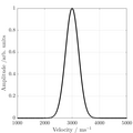
Gauss and Lorentz lineshapes.png 500 × 357; 15 KB
-
Gauss and Lorentz lineshapes.svg 1,082 × 695; 63 KB
-
Gaußprofil.jpg 612 × 452; 31 KB
-
Gr mock-up of continuous hydrogen.svg 696 × 291; 54 KB
-
Gruntman ena 01.jpg 408 × 204; 29 KB
-
Gruntman ena 02.jpg 352 × 238; 37 KB
-
H-K graph.jpg 1,813 × 1,531; 147 KB
-
Harmonique.jpg 397 × 170; 14 KB
-
HD.6C.037 (11856519893).jpg 3,200 × 2,543; 932 KB
-
High pressure sodium spectrum.png 745 × 97; 64 KB
-
HITRAN Logo.jpg 513 × 462; 99 KB
-
HITRAN Rosetta Stone.jpg 1,061 × 1,030; 377 KB
-
Holmium spectrum.png 645 × 45; 13 KB
-
Homemade Spectroscope.jpg 659 × 464; 48 KB
-
Hreels.jpg 1,071 × 565; 172 KB
-
Hyperspectral image of a copolymer blend.png 611 × 527; 400 KB
-
ICP torch.svg 550 × 1,094; 73 KB
-
Image UPS gas.jpg 5,184 × 3,456; 2.23 MB
-
Important electronic transitions in organic compounds.png 5,692 × 3,200; 138 KB
-
IMS small.gif 1,181 × 531; 92 KB
-
InfraTec-Gassensor-NDIR-Messprinzip-Abb7a.jpg 2,422 × 946; 175 KB
-
InfraTec-Gassensor-NDIR-Messprinzip-Abb7b.jpg 2,481 × 1,005; 263 KB
-
InfraTec-Gassensor-NDIR-Prinzip-Abb2.jpg 2,245 × 769; 119 KB
-
InfraTec-Gassensor-Planare-Detektoren-Abb3.jpg 1,024 × 683; 224 KB
-
InfraTec-GassensorSteckbares-Modul-Abb5.jpg 1,005 × 1,005; 132 KB
-
InfraTec-Gasssensor-Diagramm-Transmission-CO2-Abb1.jpg 1,644 × 979; 375 KB
-
Initial DLTS Result.jpg 544 × 405; 61 KB
-
Intensidade dls.png 687 × 723; 13 KB
-
InterferenceGrating flat.gif 1,004 × 446; 852 KB
-
InterferenceGrating.gif 683 × 446; 827 KB
-
InterferencePattern flat.svg 891 × 399; 204 KB
-
InterferencePattern.svg 608 × 399; 219 KB
-
Ionizációs km.jpg 553 × 1,000; 120 KB
-
Ir spectroscope.jpg 1,692 × 1,830; 771 KB
-
Ir spectroscope2.jpg 2,358 × 2,448; 1.35 MB
-
IRAF splot.png 650 × 504; 18 KB
-
IRMS.png 623 × 472; 18 KB
-
Iron spectrum.png 1,102 × 84; 31 KB
-
Irspec1.jpg 1,138 × 865; 42 KB
-
Isospunkt.svg 353 × 293; 157 KB
-
Israeli stamps 1964 - Sixteenth Independence Day, B.jpg 473 × 738; 272 KB
-
Ives Stilwell molecular and atomic spectra.png 901 × 424; 61 KB
-
Ives-Stilwell second order vs first order shifts es.svg 478 × 450; 59 KB
-
Ives-Stilwell second order vs first order shifts.svg 478 × 450; 58 KB
-
Jablonski diagram hu.png 2,530 × 1,977; 443 KB
-
Jablonski energy diagram.png 3,588 × 4,018; 348 KB
-
JablonskiSimple.png 1,192 × 926; 66 KB
-
Kirchhof laws.svg 1,375 × 1,110; 122 KB
-
Krypton gas discharge lamp - National Museum of Nature and Science, Tokyo - DSC07785.jpg 4,215 × 3,140; 2.25 MB
-
Krypton-86-lamp NIST 49.jpg 300 × 450; 21 KB
-
Kwantitatieve bepaling naproxen.JPG 547 × 483; 34 KB
-
Laboratorio de Láseres y Espectroscopía.jpg 5,184 × 3,456; 2.65 MB
-
Laboratory of electron spectroscopy.jpg 4,797 × 3,206; 9.31 MB
-
Lai-Sheng Wang (cropped).jpg 806 × 806; 140 KB
-
Lai-Sheng Wang.jpg 4,032 × 3,024; 1.4 MB
-
Laser lab (36885558663).jpg 5,592 × 3,720; 4.93 MB
-
Lattice-modes-fr.png 630 × 787; 65 KB
-
Lattice-modes.png 630 × 787; 73 KB
-
Layers make-up defocusing.png 1,232 × 752; 143 KB
-
Lboz-spectra.png 547 × 546; 5 KB
-
Lead spectrum.png 612 × 90; 53 KB


