Category:Sensory Neuroscience: Hearing and speech
Jump to navigation
Jump to search
This is a hidden category used to organize media files used in b:Sensory Neuroscience: Hearing and speech.
Subcategories
This category has only the following subcategory.
U
Media in category "Sensory Neuroscience: Hearing and speech"
The following 111 files are in this category, out of 111 total.
-
16bit sine.ogg 3.0 s; 8 KB
-
6bit sine dithered.ogg 3.0 s; 29 KB
-
6bit sine truncated.ogg 3.0 s; 23 KB
-
Acoustic filters.svg 2,301 × 1,770; 57 KB
-
Anatomy of Human Ear with Cochlear Frequency Mapping.svg 674 × 519; 33 KB
-
Anatomy of the Human Ear.svg 512 × 389; 50 KB
-
Auditory Cortex Frequency Mapping.svg 800 × 500; 110 KB
-
Band-pass filter.svg 2,770 × 1,550; 42 KB
-
Basilar membrane motion.svg 600 × 225; 19 KB
-
Basilar membrane response.svg 200 × 350; 14 KB
-
Beat frequency (1000, 1400, 1800, 2200).svg 500 × 650; 107 KB
-
Beat frequency (1200, 1400, 1600, 1800).svg 500 × 650; 86 KB
-
Beat frequency (2000, 2200, 2400, 2600).svg 500 × 650; 115 KB
-
Brain Surface Gyri.SVG 1,024 × 731; 28 KB
-
Cochlea defects in mutants.png 2,196 × 2,635; 7.42 MB
-
Cochlea-crosssection.png 640 × 537; 143 KB
-
Cochlea-trace.svg 310 × 221; 17 KB
-
Cochlea.png 389 × 226; 42 KB
-
Cochleaimplantat.jpg 882 × 768; 348 KB
-
Constructive interference.svg 250 × 300; 27 KB
-
Destructive interference1.svg 250 × 300; 24 KB
-
Ductus cochlearis schema.jpg 981 × 851; 151 KB
-
Ear internal anatomy numbered.svg 441 × 378; 183 KB
-
Flattened cochlear preparation.png 855 × 1,360; 615 KB
-
Gray904.png 285 × 450; 31 KB
-
Gray909-labels.png 350 × 350; 84 KB
-
Gray910.png 365 × 500; 43 KB
-
Gray911.png 550 × 338; 38 KB
-
Gray912.png 500 × 450; 52 KB
-
Gray913.png 550 × 492; 51 KB
-
Gray915.png 600 × 500; 44 KB
-
Gray916.png 450 × 275; 13 KB
-
Gray917.png 400 × 253; 16 KB
-
Gray918.png 313 × 177; 5 KB
-
Gray919.png 342 × 500; 36 KB
-
Gray920-labels.png 500 × 365; 110 KB
-
Gray920.png 500 × 365; 46 KB
-
Gray921 ja.png 600 × 469; 255 KB
-
Gray921.png 600 × 469; 60 KB
-
Gray922.png 500 × 463; 56 KB
-
Gray923.png 600 × 434; 61 KB
-
Gray924.png 500 × 384; 17 KB
-
Gray924.svg 500 × 384; 1.21 MB
-
Gray926.png 600 × 417; 47 KB
-
Gray927.png 500 × 461; 43 KB
-
Gray928.png 600 × 408; 50 KB
-
Gray929.png 600 × 246; 21 KB
-
Gray930.png 500 × 281; 33 KB
-
Gray931.png 600 × 334; 43 KB
-
Gray932.png 1,018 × 716; 1 MB
-
Gray933.png 400 × 288; 56 KB
-
Hair Cell Patterning Defects in the Cochlea.png 2,012 × 2,475; 3.05 MB
-
Harmonic partials on strings.svg 620 × 590; 10 KB
-
Harmonicos cordas.png 370 × 237; 8 KB
-
Harmonics of 1.2 kHz.svg 200 × 350; 13 KB
-
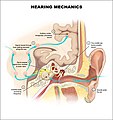
Hearing mechanics cropped - Acoustic radiation.jpg 574 × 489; 176 KB
-
Hearing mechanics cropped A1.jpg 574 × 489; 176 KB
-
Hearing mechanics cropped.jpg 574 × 489; 189 KB
-
Hearing mechanics.jpg 614 × 650; 249 KB
-
Hearing Science brainteaser - constructive interference.svg 325 × 175; 12 KB
-
High-frequency conductive hearing loss.svg 250 × 200; 12 KB
-
Human Auditory System.webm 10 min 52 s, 1,920 × 1,080; 48.32 MB
-
Huygens.gif 150 × 140; 60 KB
-
ILD vs firing rate.svg 691 × 473; 65 KB
-
Illu auditory ossicles-bn.svg 268 × 209; 89 KB
-
Illu auditory ossicles-en.svg 268 × 209; 89 KB
-
Illu auditory ossicles-es.svg 268 × 209; 89 KB
-
Illu auditory ossicles-hi.svg 268 × 209; 89 KB
-
Illu auditory ossicles-pt.svg 268 × 209; 89 KB
-
Illu auditory ossicles-ta.svg 268 × 209; 85 KB
-
Illu auditory ossicles-te.svg 268 × 209; 85 KB
-
Inner ear pathology in MPS IIIB mice at 30 wks-A.png 1,985 × 1,112; 1.18 MB
-
Inverse square law.svg 480 × 320; 5 KB
-
IPD vs firing rate.svg 600 × 500; 17 KB
-
IPD-ILD vs azimuth.svg 600 × 350; 16 KB
-
Low-frequency conductive hearing loss.svg 250 × 200; 12 KB
-
LSO wiring.svg 1,025 × 625; 108 KB
-
Mellomore.svg 189 × 178; 22 KB
-
MSO neuron wiring.svg 622 × 424; 64 KB
-
Normales Trommelfell.jpg 602 × 555; 121 KB
-
Onde stationnaire pression tuyau ferme trois modes.svg 250 × 201; 16 KB
-
Onde stationnaire pression tuyau ouvert trois modes.svg 248 × 201; 25 KB
-
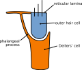
Outer hair cell and Deiter's cell.svg 274 × 233; 59 KB
-
Owl sound localization parallel processing in brain.jpg 550 × 600; 52 KB
-
Partial transmittance.gif 367 × 161; 67 KB
-
Predictions of Tuning Characteristics.svg 2,790 × 3,146; 1.89 MB
-
Psychoacoustical tuning curves.svg 400 × 275; 25 KB
-
Schematic uncoiled cochlea.svg 1,209 × 332; 33 KB
-
Sine waves different frequencies.png 480 × 100; 11 KB
-
Snail shell.jpg 2,816 × 2,112; 3.11 MB
-
Spectrogram -iua-.png 946 × 705; 300 KB
-
Spectrogram of I owe you.png 436 × 227; 70 KB
-
Standing wave.gif 750 × 250; 80 KB
-
Stapes human ear.jpg 1,236 × 927; 211 KB
-
Stapes Laserschuss2.ogv 12 s, 720 × 576; 707 KB
-
Stehende welle 3.gif 320 × 240; 238 KB
-
Stria vascularis1.jpg 1,057 × 1,078; 164 KB
-
Suppress fundamental.ogg 9.6 s; 59 KB
-
The kinocilia is mislocalized in Tailchaser hair cells.png 2,989 × 2,746; 6.19 MB
-
The Tailchaser mutation does not affect formation of interstereocilial links.png 2,756 × 2,792; 6.41 MB
-
Trommelfell.png 649 × 736; 34 KB
-
Tuning curve of OHC front row loss and IHC loss.svg 250 × 325; 11 KB
-
Tuning curve.svg 250 × 200; 9 KB
-
Two views of cochlear mechanics (A).svg 600 × 400; 38 KB
-
Two views of cochlear mechanics (B).svg 600 × 400; 51 KB
-
Two Views of Cochlear Mechanics.svg 600 × 800; 74 KB
-
Tympanic membrane cross-section.svg 285 × 203; 17 KB
-
Uncoiled cochlea with basilar membrane.png 3,487 × 2,082; 1.24 MB
-
Wave period.gif 400 × 200; 72 KB


