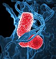
Category:Scanning electron microscopic images of bacteria
Jump to navigation
Jump to search
Subcategories
This category has the following 4 subcategories, out of 4 total.
Media in category "Scanning electron microscopic images of bacteria"
The following 77 files are in this category, out of 77 total.
-
110614 HMDS Bakterien 2 corrected.tif 2,560 × 2,048; 15.02 MB
-
Acidimicrobium ferrooxidans.jpg 698 × 469; 185 KB
-
Acinetobacter baumannii, SEM, 9330 lores.JPG 1,200 × 815; 122 KB
-
Acinetobacter baumannii.JPG 2,835 × 1,927; 525 KB
-
Algae and bacteria in Scanning Electron Microscope, magnification 2000x.JPG 1,280 × 1,024; 851 KB
-
Bacillus odysseyi.jpg 503 × 309; 26 KB
-
Bacillus subtilis R0179.jpg 457 × 552; 59 KB
-
Bacteria-driven hybrid microswimmers with a spherical body.jpg 1,000 × 589; 113 KB
-
Bordetella bronchiseptica 01.jpg 2,838 × 1,893; 2.12 MB
-
Bordetella bronchiseptica.jpg 388 × 232; 18 KB
-
Burkholderia cepacia.jpg 2,100 × 1,332; 990 KB
-
Campylobacter jejuni 01.jpg 1,500 × 1,020; 68 KB
-
Campylobacter jejuni 5778 lores.jpg 700 × 476; 48 KB
-
Cholera bacteria SEM.jpg 1,228 × 960; 343 KB
-
Citrobacter freundii.jpg 700 × 462; 62 KB
-
Clostridium difficile 01.jpg 2,835 × 1,927; 696 KB
-
Clostridium difficile EM.png 700 × 475; 218 KB
-
ClostridiumDifficile.jpg 562 × 394; 75 KB
-
Coli3.jpg 1,024 × 720; 192 KB
-
Diverse e Coli.png 712 × 514; 449 KB
-
E. coli Bacteria (1657874457).jpg 524 × 403; 70 KB
-
Ecoli dividing.jpg 2,835 × 1,927; 386 KB
-
EColiCRIS051-Fig2.jpg 760 × 384; 132 KB
-
Entercoccus sp2 lores.jpg 652 × 499; 42 KB
-
Enterococcus faecalis SEM 01 Detail.png 441 × 378; 236 KB
-
Enterococcus faecalis SEM 01.png 2,838 × 1,901; 1.85 MB
-
Esch coli.jpg 700 × 475; 71 KB
-
Escherichia coli (SEM).jpg 2,835 × 1,927; 671 KB
-
Exiguobacterium sp. S17, scanning electron micrograph.png 1,069 × 602; 661 KB
-
Gemmatimonas aurantiaca.jpg 1,260 × 909; 157 KB
-
Gemmatimonas groenlandica.jpg 1,583 × 2,091; 353 KB
-
Helicobacter pylori.jpg 138 × 200; 16 KB
-
Helicobacter pylori2.jpg 138 × 200; 23 KB
-
Helicobacter sp 01.jpg 1,500 × 1,020; 802 KB
-
HelicobacterPylori2.jpg 700 × 476; 64 KB
-
Hpylori.jpg 700 × 476; 53 KB
-
Klebsiella pneumoniae Bacterium (13383411493).jpg 2,539 × 2,657; 950 KB
-
Klebsiella-pneumoniae.jpg 700 × 475; 61 KB
-
Legionella pneumophila (SEM) 2.jpg 2,835 × 1,927; 790 KB
-
Legionella pneumophila (SEM).jpg 2,835 × 1,927; 745 KB
-
LeishmaniaMexicana Promastigote SEM.jpg 1,280 × 960; 388 KB
-
Leptospira scanning micrograph.jpg 700 × 470; 110 KB
-
Methylomonas methanica EID.jpg 600 × 584; 35 KB
-
Micrococcus luteus 9756.jpeg 2,835 × 1,927; 1.82 MB
-
Micrococcus luteus 9756.tif 2,835 × 1,927; 3.98 MB
-
Micrococcus luteus 9757.jpeg 2,835 × 1,927; 1.82 MB
-
Micrococcus luteus 9758.jpeg 2,835 × 1,927; 1.77 MB
-
Micrococcus luteus 9759.jpeg 2,835 × 1,927; 1.78 MB
-
Micrococcus luteus 9760.jpeg 2,835 × 1,927; 1.64 MB
-
Micrococcus luteus 9761.jpeg 2,835 × 1,927; 1.63 MB
-
Mixed-culture biofilm.jpg 2,560 × 1,920; 3.06 MB
-
Multidrug-resistant Klebsiella pneumoniaeand neutrophil.jpg 732 × 768; 284 KB
-
Mycobacterium fortuitum.png 2,835 × 1,927; 7.69 MB
-
Pseudomonas aeruginosa SEM.jpg 2,676 × 1,879; 785 KB
-
Pseudomonas.jpg 2,676 × 1,879; 496 KB
-
Ralstonia mannitolilytica.png 2,835 × 1,927; 3.72 MB
-
Rickettsiatyphi.jpg 1,817 × 1,744; 3.91 MB
-
Ruffle Formation Induced by S. Typhimurium (8515863563).jpg 265 × 194; 26 KB
-
Salmobandeau.jpg 353 × 299; 27 KB
-
Salmonella Bacteria (5613656967).jpg 640 × 536; 56 KB
-
Salmonella enteritidis in color.jpg 1,979 × 2,250; 1.69 MB
-
Salmonella typhimurium.png 1,010 × 757; 396 KB
-
SalmonellaNIAID.jpg 2,100 × 1,761; 1.66 MB
-
Salmonelle2d.jpg 340 × 312; 25 KB
-
Shewanella oneidensis.png 648 × 377; 117 KB
-
Symbiotic nitrogen fixing bacteria inside legume root nodule cells.tif 1,024 × 1,224; 2.4 MB
-
Treponema pallidum Bacteria (Syphilis) (cropped).jpg 2,076 × 2,134; 2.37 MB
-
Treponema pallidum.jpg 531 × 500; 42 KB
-
TreponemaPallidum.jpg 572 × 500; 61 KB
-
USDAbacteria.jpg 640 × 511; 105 KB
-
Vancomycin-Resistant Enterococcus 01.jpg 2,838 × 1,910; 405 KB
-
Venenivibrio.jpg 478 × 653; 254 KB
-
Vibrio parahaemolyticus 01.jpg 2,835 × 1,927; 2.32 MB
-
Vibrio vulnificus 01.png 629 × 420; 530 KB


