Category:Scanning electron microscopic images of animals
Jump to navigation
Jump to search
Subcategories
This category has the following 4 subcategories, out of 4 total.
Media in category "Scanning electron microscopic images of animals"
The following 86 files are in this category, out of 86 total.
-
Antonae guttipes REM.jpg 1,890 × 1,413; 1.4 MB
-
Bdelloid.JPG 1,417 × 1,483; 238 KB
-
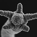
Brachiolarian arms.JPG 1,024 × 976; 366 KB
-
Cabeza de milpiés vista bajo el microscopio electrónico de barrido de electrones.jpg 3,072 × 2,094; 4.35 MB
-
Cabeza de ácaro trombidiforme en vista dorsal bajo el microscopio electrónico de barrido.jpg 3,072 × 2,094; 4.16 MB
-
Comparison of dentary teeth of Oromycter and Arisierpeton SEM images.png 2,549 × 1,002; 1.4 MB
-
Cruz-Lacierda et al Pseudorhabdosynochus AACLBioflux 2012.png 1,595 × 1,631; 697 KB
-
Crystal formations? (3501294903).jpg 1,280 × 960; 281 KB
-
CSIRO ScienceImage 1391 Scanning Electron Micrograph of an Oyster Spat.jpg 2,657 × 2,193; 4.82 MB
-
DDC-SEM of fossil - green - 1.jpg 1,024 × 708; 993 KB
-
DDC-SEM of fossil - green - 2.jpg 1,024 × 710; 992 KB
-
DDC-SEM of fossil - orange - 1.jpg 1,024 × 704; 857 KB
-
DDC-SEM of fossil - orange - 2.jpg 1,024 × 706; 980 KB
-
DDC-SEM of fossil - pink - 1.jpg 1,024 × 710; 964 KB
-
DDC-SEM of fossil - purple - 1.jpg 1,024 × 710; 841 KB
-
DDC-SEM of fossil - purple - 2.jpg 1,024 × 708; 826 KB
-
DDC-SEM of fossil - purple - 3.jpg 1,024 × 710; 993 KB
-
DDC-SEM of fossil - red - 1.jpg 1,024 × 710; 773 KB
-
Eponge.jpg 1,024 × 1,106; 448 KB
-
Feather 2.jpg 600 × 432; 83 KB
-
Fenestrulina delicia.tif 1,280 × 1,080; 1.33 MB
-
Geckofot edited.png 639 × 1,445; 1.16 MB
-
Geckofot.jpg 1,920 × 480; 238 KB
-
Haircell frog sacculus.jpg 391 × 334; 639 KB
-
HederelloidSEM.jpg 457 × 383; 69 KB
-
Hemipristis elongatus anterior (3501296073).jpg 1,519 × 829; 478 KB
-
Hemipristis serra lateral (3502111018).jpg 1,280 × 960; 144 KB
-
Hydra magnipapillata.jpg 400 × 400; 18 KB
-
Larva on scanning electron microscope.jpg 1,220 × 1,024; 1.66 MB
-
Oromycter and Arisierpeton, second premaxillary tooth comparison.png 1,398 × 1,150; 745 KB
-
Parasite130103-fig2 Protopolystoma xenopodis (Monogenea, Polystomatidae) Oncomiracidium.tif 2,067 × 2,027; 4.81 MB
-
Parasite130103-fig4 Protopolystoma xenopodis (Monogenea, Polystomatidae) Adult.tif 2,067 × 2,044; 2.24 MB
-
Parasite140015-fig1 Protoopalina pingi (Opalinidae) SEM.tif 2,343 × 1,758; 1.59 MB
-
Parasite140076-fig1 Dirofilaria repens removed from a subcutaneous nodule - Photos.png 1,645 × 2,894; 5.31 MB
-
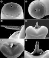
Parasite140131-fig2 Capillaria plectropomi (Nematoda) - Scannin Electron Microscopy.tif 2,835 × 3,402, 2 pages; 7.15 MB
-
Parasite140132-fig2 Philometra protonibeae (Nematoda, Philometridae).png 2,067 × 2,480; 3.09 MB
-
Parasite160002-fig2 Philometra inexpectata.tif 2,835 × 3,508; 6.59 MB
-
Parasite160002-fig4 Philometra jordanoi.tiff 2,835 × 3,596; 6.86 MB
-
Parasite160108-fig3 - Triloculotrema euzeti (Monogenea, Monocotylidae).png 1,417 × 1,350; 1.64 MB
-
Parasite170054-fig2 Rhadinorhynchus oligospinosus (Acanthocephala).png 2,657 × 3,092; 3.4 MB
-
Parasite170054-fig3 Rhadinorhynchus oligospinosus (Acanthocephala).png 2,657 × 3,122; 4.13 MB
-
Parasite170054-fig4 Rhadinorhynchus oligospinosus (Acanthocephala).png 2,657 × 3,120; 12.05 MB
-
Parasite180037-fig5 FIGS 15-21 Pararhadinorhynchus magnus.png 2,864 × 3,720; 4.99 MB
-
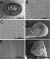
Parasite180056-fig2A Placobdelloides siamensis (Glossiphoniidae).png 1,024 × 882; 1 MB
-
Parasite180056-fig2B Placobdelloides siamensis (Glossiphoniidae).png 1,600 × 1,152; 1.43 MB
-
Parasite180056-fig3 Placobdelloides siamensis (Glossiphoniidae).png 1,024 × 877; 949 KB
-
Parasite180057-fig1 Chloromyxum atlantoraji SEM.png 1,772 × 1,961; 2.36 MB
-
Parasite180057-fig3 Chloromyxum zearaji SEM.png 2,008 × 1,045; 1.47 MB
-
Parasite180057-fig5 Chloromyxum riorajum SEM.png 1,654 × 1,248; 866 KB
-
Parasite180070-fig3 Rasheedia heptacanthi (Nematoda, Physalopteridae).png 3,189 × 3,828; 5.49 MB
-
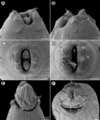
Parasite180099-fig2 Cucullanus austropacificus (Nematoda, Cucullanidae).png 1,417 × 2,764; 2.35 MB
-
Parasite180099-fig3 Cucullanus austropacificus (Nematoda, Cucullanidae).png 2,835 × 2,835; 2.63 MB
-
Parasite180099-fig5 Cucullanus gymnothoracis (Nematoda, Cucullanidae).png 2,835 × 3,402; 4.32 MB
-
Parasite180099-fig6 Cucullanus gymnothoracis (Nematoda, Cucullanidae).png 2,835 × 2,268; 3 MB
-
Parasite180099-fig8 Cucullanus incognitus (Nematoda, Cucullanidae).png 2,835 × 3,685; 3.64 MB
-
PLoS Rotaria.gif 1,949 × 4,103; 1.88 MB
-
Polychaeta. Colored SEM Image.png 5,945 × 5,945; 132.04 MB
-
Polychaeta. SEM Image.png 5,945 × 5,945; 45.13 MB
-
Salaria fluviatilis SEM imaging.jpg 1,573 × 4,079; 3.96 MB
-
Schistosome Parasite SEM.jpg 1,800 × 2,224; 922 KB
-
SEM Carcharhinus leucas, 9x (3261812961).jpg 1,116 × 960; 313 KB
-
SEM Hemipristis serra tip, 9x (3261812869).jpg 1,280 × 960; 198 KB
-
SEM Hemipristis serra, 9x (3262640074).jpg 1,280 × 960; 205 KB
-
SEM image of Milnesium tardigradum in active state - journal.pone.0045682.g001-2.png 1,572 × 1,205; 3.05 MB
-
SEM image of Milnesium tardigradum in tun state - journal.pone.0045682.g001-3.png 1,507 × 1,176; 1.75 MB
-
SEM photo of S. mediterranea.jpg 1,280 × 1,024; 440 KB
-
Shell surface.tif 1,280 × 1,040; 1.27 MB
-
Shell-unknown3 hg.jpg 2,820 × 2,190; 1.37 MB
-
Shell-unknown5 hg.jpg 2,880 × 2,262; 1.31 MB
-
So4b-08.jpg 712 × 484; 224 KB
-
Soybean cyst nematode and egg SEM.jpg 1,944 × 1,599; 970 KB
-
Steinernema carpocapsae SEM.tif 1,424 × 1,064; 1.45 MB
-
Stereocilia of frog inner ear.01.jpg 438 × 311; 38 KB
-
Зуб скумбрии в электронном микроскопе.jpg 1,502 × 1,533; 2.45 MB


