Category:Scanning electron microscopic images
Jump to navigation
Jump to search
Italiano: microscopio elettronico a scansione (SEM)
Subcategories
This category has the following 14 subcategories, out of 14 total.
M
S
Media in category "Scanning electron microscopic images"
The following 200 files are in this category, out of 393 total.
(previous page) (next page)-
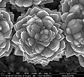
"Цветок". Макрофото нанотрубок.jpg 1,024 × 943; 630 KB
-
"Ящер". Макрофото нанотрубок.jpg 4,096 × 3,773; 1.25 MB
-
1 calcified carotid.jpg 1,019 × 658; 182 KB
-
11166 lores.jpg 700 × 475; 50 KB
-
12915 2020 762 Fig11 Pigoraptor chileana.png 1,946 × 716; 644 KB
-
12915 2020 762 Fig2-Syssomonas multiformis.webp 1,946 × 1,772; 450 KB
-
1516-1439-mr-1980-5373-MR-2016-0210-gf02.jpg 2,502 × 957; 205 KB
-
1979-rem 01 hg.jpg 2,770 × 2,773; 1.73 MB
-
1979-rem 02 hg.jpg 3,444 × 2,999; 2.83 MB
-
1979-rem 03 hg.jpg 2,734 × 1,947; 1.06 MB
-
1979-rem 04 hg.jpg 2,200 × 2,226; 1.29 MB
-
1979-rem 05 hg.jpg 1,870 × 1,822; 1.05 MB
-
1979-rem 06 hg.jpg 1,856 × 1,862; 981 KB
-
1979-rem 07 hg.jpg 2,720 × 1,955; 1.09 MB
-
1979-rem 08 hg.jpg 2,692 × 1,944; 1,015 KB
-
1979-rem 09 hg.jpg 2,692 × 1,932; 1.11 MB
-
1979-rem 10 hg.jpg 2,692 × 1,950; 1.01 MB
-
1979-rem 11 hg.jpg 2,692 × 1,950; 1.4 MB
-
1979-rem 12 hg.jpg 3,460 × 2,677; 2.1 MB
-
1979-rem 13 hg.jpg 3,452 × 2,798; 1.78 MB
-
1979-rem 14 hg.jpg 2,740 × 2,739; 1.96 MB
-
1zzzzz.jpg 345 × 450; 48 KB
-
2 calcified carotid.jpg 992 × 692; 823 KB
-
253 2019 9846 Fig4.webp 2,055 × 2,760; 872 KB
-
253 2019 9846 Fig4l.jpg 761 × 2,753; 1.04 MB
-
253 2019 9846 Fig4lt.jpg 757 × 503; 222 KB
-
2artdeco (5615551876).jpg 2,048 × 2,240; 737 KB
-
2bigcrystal (5614973921).jpg 2,048 × 2,240; 1 MB
-
2calcium (5615557428).jpg 2,048 × 2,240; 487 KB
-
2catclaw (5614970985).jpg 2,048 × 2,240; 1.01 MB
-
2coin (5615555742).jpg 2,048 × 2,240; 759 KB
-
2fingernail (5615553792).jpg 2,048 × 2,240; 598 KB
-
2flower (5614967529).jpg 2,048 × 2,240; 995 KB
-
2floweroffcenter (5614968607).jpg 2,048 × 2,240; 1.03 MB
-
2flowerpetal740 (5615548400).jpg 2,048 × 2,240; 1.15 MB
-
2francis (5614970371).jpg 2,048 × 2,240; 1.36 MB
-
2glass1200 (5615549524).jpg 2,048 × 2,240; 1.49 MB
-
2jupiter (5615554548).jpg 2,048 × 2,240; 1.38 MB
-
2mica (5614974965).jpg 2,048 × 2,240; 807 KB
-
2paint (5615556328).jpg 2,048 × 2,240; 1.64 MB
-
2paint2 (5614976683).jpg 2,048 × 2,240; 1.46 MB
-
2shell2000 (5614967181).jpg 2,048 × 2,240; 469 KB
-
3zzzzz.jpg 271 × 450; 52 KB
-
40793 2015 2030327 Fig2.webp 696 × 529; 64 KB
-
40793 2017 296 Fig2.webp 473 × 470; 35 KB
-
41467 2016 Article BFncomms10543 Fig1c(top) HTML.png 244 × 153; 38 KB
-
41467 2020 20149 Fig1b.jpg 608 × 395; 79 KB
-
41467 2022 28065 Fig1a.jpg 628 × 471; 102 KB
-
41467 2022 28065 Fig1d.jpg 629 × 471; 116 KB
-
41467 2023 40657 Fig1e.jpg 860 × 650; 119 KB
-
41586 2021 3297 Fig1a-d.jpg 888 × 1,124; 225 KB
-
41598 2023 29969 Fig10 Coolia malayensis.webp 1,770 × 2,291; 633 KB
-
41598 2023 29969 Fig10GK Coolia malayensis SEM.jpg 641 × 1,049; 176 KB
-
41598 2023 29969 Fig11 Coolia malayensis.webp 1,498 × 570; 152 KB
-
41598 2023 29969 Fig11A-Coolia malayensis SEM.jpg 790 × 570; 128 KB
-
41598 2023 29969 Fig3 Ostreopsis tairoto.webp 1,498 × 1,816; 414 KB
-
41598 2023 29969 Fig3CE-Ostreopsis tairoto SEM.jpg 697 × 708; 164 KB
-
41598 2023 29969 Fig4 Ostreopsis tairoto.webp 1,770 × 1,754; 565 KB
-
41598 2023 29969 Fig6 Ostreopsis lenticularis.webp 1,498 × 2,056; 549 KB
-
41598 2023 29969 Fig6D Ostreopsis lenticularis SEM.jpg 887 × 1,343; 315 KB
-
41598 2023 29969 Fig7 Ostreopsis lenticularis.webp 1,498 × 1,752; 386 KB
-
41598 2023 29969 Fig7B Ostreopsis lenticularis-SEM.jpg 762 × 958; 207 KB
-
41598 2023 29969 Fig8 Ostreopsis lenticularis.webp 1,498 × 1,109; 452 KB
-
41598 2023 29969 Fig8B Ostreopsis lenticularis SEM.jpg 961 × 430; 165 KB
-
41598 2023 29969 Fig8D Ostreopsis lenticularis SEM.jpg 674 × 670; 175 KB
-
4zzzzz.jpg 450 × 433; 79 KB
-
5zzzzz.jpg 292 × 450; 47 KB
-
Acanthamoeba polyphaga PHIL11892.tif 2,835 × 1,927; 5.36 MB
-
AZM Euplotes vanleeuwenhoeki.tif 1,494 × 1,996; 2.02 MB
-
Back of the abdomen Aphid.tif 3,072 × 2,304; 6.76 MB
-
Braarudosphaera bigelowii.png 2,140 × 1,434; 4.48 MB
-
Cancerdusein3 DCIS Wiki.jpg 533 × 373; 119 KB
-
Cellular Uptake NPs.jpg 1,536 × 1,103; 2.11 MB
-
Cellules de liège.png 787 × 538; 279 KB
-
Chip-out of film.jpg 1,087 × 1,001; 287 KB
-
Chlamydospores - Reproductive Structure of the Phytophthora.png 1,393 × 901; 236 KB
-
Color SEM 5.jpg 2,014 × 1,345; 1.11 MB
-
Colponema vietnamica 4A pone.0095467.tif 997 × 2,273; 1.91 MB
-
Compound flower with pollen no scale bar.jpg 600 × 430; 87 KB
-
Conductive probe SEM images.png 964 × 263; 133 KB
-
Copper plated Heterojunction solar cell SEM-EDS image.png 1,954 × 1,085; 5.34 MB
-
Corrosion T-rex grows from the steel bar in reinforced concrete.pdf 1,566 × 1,441; 2.91 MB
-
Coscinodiscus oculus-iridis (Ehrenberg) Ehrenberg 1840 diatom shell. Colored SEM image.jpg 4,500 × 4,500; 18.8 MB
-
Coscinodiscus oculus-iridis (Ehrenberg) Ehrenberg 1840 diatom shell. Colored SEM image.png 4,500 × 4,500; 44.33 MB
-
Coscinodiscus oculus-iridis (Ehrenberg) Ehrenberg 1840 diatom shell. SEM image.png 4,500 × 4,500; 19.41 MB
-
Crystal wings.png 689 × 690; 461 KB
-
CSIRO ScienceImage 293 Cells Interacting With Collagen.jpg 2,520 × 1,709; 3.79 MB
-
CSIRO ScienceImage 6690 SEM dinoflagellate.jpg 1,863 × 1,220; 1.72 MB
-
CSIRO ScienceImage 6743 SEM Cryptophyte.jpg 1,482 × 1,202; 1.95 MB
-
CSIRO ScienceImage 6745 derwent sample.jpg 1,863 × 1,220; 1.84 MB
-
CSIRO ScienceImage 7232 Nylon fibre.jpg 993 × 710; 354 KB
-
CSIRO ScienceImage 8440 Polyester fibre.jpg 990 × 477; 258 KB
-
Ctenorillo meyeri Taiti, 2018.jpg 1,024 × 943; 100 KB
-
Curcumin Liposomes (15000X).tif 2,560 × 1,920; 4.69 MB
-
DDC-SEM of fossil - blue - 1.jpg 1,024 × 708; 948 KB
-
DDC-SEM of fossil - blue - 2.jpg 1,024 × 710; 991 KB
-
Dental pulp stem cells. SEM-BSE.jpg 1,536 × 1,152; 1.08 MB
-
Departamentodemateriales.jpg 152 × 162; 28 KB
-
Detalle de dorso de cochinilla harinosa de la familia pseudococcidae.jpg 2,048 × 1,392; 1.5 MB
-
Detalle de polen de girasol y cardo visto bajo el microscopio electrónico de barrido.tif 2,048 × 1,536; 3.1 MB
-
Diabase (PYRS-84-3).jpg 1,500 × 1,052; 665 KB
-
Diatoms from moss.png 3,080 × 1,888; 3.35 MB
-
Dinoflagellates.jpg 480 × 330; 61 KB
-
Discoaster-diagenese rem hg.jpg 2,560 × 1,920; 3.91 MB
-
Dysderocrates silvestris 05 b.tif 856 × 888; 742 KB
-
Emiluvia premyogii.png 699 × 464; 60 KB
-
Epidermis of Festuca arundinacea.jpg 8,499 × 6,178; 9.12 MB
-
Epistominella-exigua hg.jpg 2,582 × 2,204; 1.12 MB
-
ESEM color salt hydrationx.png 2,322 × 814; 1.89 MB
-
Evolutionary Movement.jpg 1,280 × 960; 135 KB
-
F-urine 02 50x BED.jpg 1,023 × 762; 1.26 MB
-
Fern leave like crystals formed in glass-ceramics.jpg 1,024 × 864; 270 KB
-
Fibers x60.tif 2,560 × 1,920; 4.69 MB
-
Fig1 MEB C2.tif 720 × 960; 399 KB
-
Fig1-Scanning-electron-microscope-SEM-for-graphene-oxide-GO.jpg 388 × 292; 20 KB
-
Figure-3-SEM-micrograph-of-Ag-microclusters-covering-a-silicon-grain.jpg 1,200 × 1,140; 276 KB
-
Filière d' Hahnia.jpg 2,923 × 2,425; 828 KB
-
Filter paper 840 3x3 copy.jpg 4,800 × 4,000; 1.63 MB
-
Flowerette in a Composite Flower.jpg 6,096 × 4,485; 3.08 MB
-
Fotonic fiber x6000.JPG 1,280 × 960; 181 KB
-
Funktionsprinzip REM.gif 976 × 560; 31 KB
-
Fusitriton oregonensis parasperm.png 3,038 × 1,571; 475 KB
-
Fusules de Micrathena.jpg 3,483 × 2,490; 749 KB
-
Fusules de Nesticus.jpg 3,471 × 2,557; 811 KB
-
Fusules de Scytodes.jpg 3,931 × 2,773; 846 KB
-
Gamma-prime FOV 10.png 3,072 × 3,072; 5.92 MB
-
Gamma-prime FOV 4um.png 3,072 × 3,072; 5.87 MB
-
Gamma-Ray SEM Jeong et al.png 476 × 208; 96 KB
-
GO SEM.png 840 × 341; 314 KB
-
Gongylonema pulchrum mouth.tif 572 × 665; 516 KB
-
Granat zoniert.jpg 1,281 × 1,024; 178 KB
-
Graphite Pencil.JPG 1,024 × 720; 134 KB
-
Halobacterium salinarum NRC-1.png 600 × 740; 136 KB
-
Halomonas sp. R5-57.png 472 × 312; 100 KB
-
Harmannella entrapping Legionella.png 972 × 1,172; 1.92 MB
-
Hartmannella vermiformis.jpg 2,835 × 1,927; 540 KB
-
Hemieuryale pustulata (10.11646-zootaxa.3925.3.2) Figure 3.png 1,866 × 2,630; 6.34 MB
-
Hoar crystal usda darling-wolf.jpg 768 × 1,024; 152 KB
-
Homeostasis del eritrocito y la hemoglobina.png 1,361 × 1,814; 918 KB
-
Human corneal epithelium. SEM-BSE.jpg 3,072 × 2,304; 3.24 MB
-
Human Respiratory Syncytial Virus (RSV) - 52453988775.jpg 2,048 × 1,536; 1.82 MB
-
Ichthyophthirius multifiliis theront.tif 2,417 × 1,249; 4.59 MB
-
InGaN crystal SEM+CL.png 1,298 × 928; 1.73 MB
-
Insulin green microtrees.jpg 582 × 450; 111 KB
-
Insulin microtrees.png 1,024 × 713; 561 KB
-
Intergranular corrosion.JPG 1,348 × 1,052; 171 KB
-
IRG activation following pathogen entry.jpg 949 × 725; 156 KB
-
Jmse-11-00001-g014.webp 3,870 × 1,095; 112 KB
-
Jmse-11-00001-g014C.jpg 238 × 116; 8 KB
-
Karenia brevis.jpg 379 × 383; 16 KB
-
La0.2Ca0.8MnO3.png 378 × 630; 73 KB
-
Lagenid microstructure.png 1,238 × 928; 736 KB
-
Lame maxillaire de Leptyphantes.jpg 3,337 × 2,551; 1.02 MB
-
Layered yttrium hydroxide.png 2,048 × 1,398; 9.56 MB
-
LeishmaniaMexicana Amastigote SEM.jpg 1,280 × 960; 469 KB
-
Life Science Imaging with ZEISS Scanning Electron Microscopes (10690272504).jpg 5,614 × 6,876; 3.9 MB
-
Limbal epithelial cells. Monolayer. SEM-BSE.jpg 3,072 × 2,304; 4.74 MB
-
Limbal epithelial cells. SEM-BSE.jpg 3,072 × 2,304; 4.93 MB
-
Limbal epithelium. Cells. SEM-BSE.jpg 3,072 × 2,304; 4.31 MB
-
Macs killing cancer cell.jpg 2,289 × 1,669; 1.08 MB
-
Magnetic Beads Spider.tif 3,072 × 2,045; 5.57 MB
-
Malassezia lipophilis 3 lores.jpg 700 × 497; 27 KB
-
Mallomonas punctifera.tif 5,120 × 3,840; 18.77 MB
-
Marr micrograph.tiff 1,280 × 1,100; 1.35 MB
-
Micrometric coral and fish.tif 2,048 × 1,756; 10.29 MB
-
Micron Sized Cat - Meoww.tif 1,024 × 768; 1.89 MB
-
Miliolid wall SEM.png 886 × 570; 470 KB
-
Moravec & Justine - Euterranova n. gen. and Neoterranova n. gen - parasite200141-fig2.png 3,484 × 4,179; 4.98 MB
-
Moravec & Justine - Euterranova n. gen. and Neoterranova n. gen - parasite200141-fig3.png 3,484 × 4,179; 6.26 MB
-
Moravec & Justine - Euterranova n. gen. and Neoterranova n. gen - parasite200141-fig5.png 3,484 × 2,788; 4.64 MB
-
My 10 micrometer heart.tif 1,024 × 888; 897 KB
-
Mycelial film.jpg 1,280 × 960; 449 KB
-
Nano-Explosion.jpg 4,942 × 3,249; 7.97 MB
-
Nano-particle Christmas Tree.jpg 3,850 × 2,850; 6.9 MB
-
Nanocristales vistos bajo el microscopio electrónico de barrido.jpg 2,048 × 1,392; 1.89 MB
-
Nanoimprinted-campanile-probe.png 1,412 × 833; 1.6 MB
-
Nanoplankton-fossil-sediment hg.jpg 2,841 × 2,244; 1.29 MB
-
Nanowire; Practical Oscillators (5941044200).jpg 365 × 314; 130 KB
-
Nebela flabellulum SEM.png 841 × 1,052; 103 KB
-
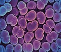
Neisseria gonorrhoeae Bacteria - 53136008302.jpg 2,048 × 1,752; 3.19 MB
-
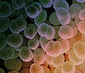
Neisseria gonorrhoeae Bacteria - 53136604826.jpg 2,048 × 1,740; 3.11 MB
-
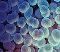
Neisseria gonorrhoeae Bacteria - 53136805124.jpg 3,600 × 3,094; 3.12 MB
-
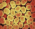
Neisseria gonorrhoeae Bacteria - 53137010205.jpg 3,600 × 3,094; 3.65 MB
-
Neisseria gonorrhoeae Bacteria.jpg 2,048 × 1,740; 3.56 MB
-
Neisseria meningitidis Charles-Orszag 2018.png 1,433 × 1,433; 2.25 MB
-
Neurologic Main.png 738 × 515; 328 KB
-
Neuron-SEM-2.png 800 × 600; 114 KB
-
Nodal cilia.jpg 560 × 372; 56 KB
-
Notocera spinidorsa REM.jpg 1,890 × 1,410; 1.44 MB
