Category:Ribbon diagrams of lyases
Jump to navigation
Jump to search
Media in category "Ribbon diagrams of lyases"
The following 129 files are in this category, out of 129 total.
-
 1a5c.jpg 240 × 240; 28 KB
-
 1aa1.jpg 1,000 × 794; 302 KB
-
 1aos.jpg 1,000 × 797; 242 KB
-
 1b66.jpg 1,000 × 779; 237 KB
-
 1ca2.jpg 240 × 240; 24 KB
-
 1d7k.jpg 1,000 × 797; 320 KB
-
 1dw9.jpg 1,000 × 797; 296 KB
-
 1dw92.jpg 1,000 × 797; 383 KB
-
 1e51.jpg 1,000 × 794; 345 KB
-
 1ecq.jpg 1,000 × 797; 404 KB
-
 1ef8.jpg 1,000 × 794; 286 KB
-
 1fuo.jpg 1,000 × 794; 342 KB
-
 1fuo2.jpg 1,553 × 1,000; 1.04 MB
-
 1gkm.jpg 1,000 × 797; 354 KB
-
 1hrk.jpg 1,000 × 793; 316 KB
-
 1i7q.jpg 1,000 × 797; 334 KB
-
 1j2y.jpg 1,000 × 797; 293 KB
-
 1jbq.jpg 1,000 × 797; 326 KB
-
 1jr2.jpg 240 × 240; 9 KB
-
 1lk9.jpg 240 × 240; 21 KB
-
 1lw4.jpg 1,000 × 780; 348 KB
-
 1nhx.jpg 1,000 × 792; 288 KB
-
 1nrx.jpg 1,100 × 800; 349 KB
-
 1nvm.jpg 1,435 × 792; 352 KB
-
 1one.jpg 1,000 × 794; 327 KB
-
 1p3y.jpg 1,000 × 797; 344 KB
-
 1p5j.jpg 1,000 × 797; 227 KB
-
 1pk0.jpg 1,000 × 797; 343 KB
-
 1qb4.jpg 1,000 × 797; 345 KB
-
 1qxo.jpg 1,000 × 800; 417 KB
-
 1sgj.jpg 1,000 × 794; 215 KB
-
 1t6m.png 801 × 743; 76 KB
-
 1tdj.jpg 1,000 × 797; 291 KB
-
 2b69.jpg 1,000 × 794; 246 KB
-
 2cw6.jpg 1,000 × 797; 272 KB
-
 2eb5.jpg 1,000 × 800; 344 KB
-
 2fkn.jpg 1,000 × 797; 291 KB
-
 2h31.jpg 1,000 × 794; 324 KB
-
 2h31a.jpg 1,000 × 794; 336 KB
-
 2j91.jpg 1,000 × 797; 331 KB
-
 2jib.jpg 1,000 × 793; 358 KB
-
 2jis.jpg 1,100 × 780; 332 KB
-
 2nmp.jpg 1,000 × 794; 381 KB
-
 2pan.jpg 1,000 × 792; 394 KB
-
 2pfd.jpg 1,000 × 779; 371 KB
-
 2pfd2.jpg 1,000 × 779; 441 KB
-
 2wm1.jpg 1,000 × 797; 263 KB
-
 2wqt.jpg 1,000 × 800; 445 KB
-
 2wqt2.jpg 1,000 × 792; 420 KB
-
 2ygw.png 467 × 324; 130 KB
-
 3C16 (GNAS AC5 AC2 ATP Forskolin).png 858 × 923; 373 KB
-
 3c8w.jpg 1,000 × 794; 350 KB
-
 3d8n.jpg 1,000 × 797; 201 KB
-
 3fdd.jpg 1,000 × 797; 295 KB
-
 3fmx.jpg 1,000 × 797; 350 KB
-
 3fmx2.jpg 1,000 × 797; 373 KB
-
 3leo.jpg 1,000 × 797; 224 KB
-
 3mwb.jpg 1,435 × 800; 438 KB
-
 3n75.jpg 1,000 × 800; 396 KB
-
 3nz4.jpg 1,000 × 797; 267 KB
-
 3r6v.jpg 1,000 × 793; 337 KB
-
 3rbf.jpg 1,000 × 797; 282 KB
-
 3tuu.jpg 1,000 × 800; 357 KB
-
 4c3t.jpg 1,000 × 797; 322 KB
-
 4crz.jpg 1,000 × 797; 277 KB
-
 4e1o.jpg 1,160 × 885; 400 KB
-
 4f0x.jpg 1,000 × 800; 296 KB
-
 4itx.jpg 1,000 × 797; 385 KB
-
 4kp2.jpg 1,400 × 800; 360 KB
-
 4l0o.jpg 1,000 × 800; 393 KB
-
 4lom.jpg 1,000 × 797; 425 KB
-
 4mzx.jpg 1,000 × 797; 360 KB
-
 4ntn.jpg 1,000 × 797; 263 KB
-
 4oma.jpg 1,000 × 797; 375 KB
-
 4qii.jpg 1,000 × 797; 321 KB
-
 4s27.jpg 1,000 × 797; 276 KB
-
 4w1y.jpg 1,000 × 797; 394 KB
-
 4y6g.jpg 1,000 × 797; 208 KB
-
 5afd.jpg 1,000 × 797; 329 KB
-
 5f3m.jpg 1,000 × 797; 296 KB
-
 5hi0.jpg 1,000 × 797; 327 KB
-
 5j04.jpg 1,000 × 797; 345 KB
-
 5jbx.jpg 1,000 × 800; 314 KB
-
 6gpj.jpg 1,000 × 797; 398 KB
-
 6k31.jpg 1,220 × 793; 410 KB
-
 6w2m.jpg 1,040 × 793; 291 KB
-
 6zmq.jpg 1,070 × 778; 232 KB
-
 7ACN.jpg 775 × 650; 463 KB
-
 ACCsynthasecomplexwPLP.png 958 × 596; 138 KB
-
 ACS & PLP complex with labeled 278 and 152 residues.png 1,067 × 627; 295 KB
-
 ACS white background.png 1,204 × 627; 174 KB
-
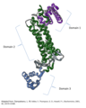 ASLMonomerwithdomains.png 518 × 650; 380 KB
-

-
 Carbonic anhydrase 1CA2 active site.png 1,240 × 1,240; 445 KB
-
 Carbonic anhydrase 1CA2.png 1,200 × 1,200; 586 KB
-
 Carbonic anhydrase.jpg 500 × 500; 125 KB
-
 Carbonic anhydrase.png 611 × 558; 60 KB
-
 DOPA decarboxylase dimer 1JS3.png 1,000 × 741; 410 KB
-
 Fumarase (yeast).jpg 500 × 500; 37 KB
-
 Fumarase.png 668 × 600; 284 KB
-
 Fumarase01.png 1,060 × 952; 564 KB
-
 GLO1 Homo sapiens small fast.gif 300 × 260; 1.11 MB
-
 GLO1 Homo sapiens.gif 560 × 400; 7.56 MB
-
 GLO1 Leishmania major small fast.gif 300 × 245; 1.13 MB
-
 GLO1 Leishmania major.gif 480 × 400; 7.43 MB
-
 HMG-CoA Lyase 2cw6.png 958 × 854; 340 KB
-
 Hyaluronidase-1OJN.png 468 × 361; 93 KB
-
 Isochorismate Pyruvate Lyase.png 661 × 439; 74 KB
-
 Lysine decarboxylase.png 2,400 × 2,400; 3.85 MB
-
 MICL.png 565 × 497; 69 KB
-
 OPRTase in Complex with OMP.jpg 500 × 500; 59 KB
-
 Ornithine Decarboxylase Publication View.png 640 × 480; 213 KB
-
 PBB Protein CBS image.jpg 500 × 500; 60 KB
-
 PBB Protein PCK1 image.jpg 500 × 500; 54 KB
-
 PDB 1fuo EBI.jpg 800 × 600; 51 KB
-
 PDB 1fup EBI.jpg 800 × 600; 51 KB
-
 PDB 1fuq EBI.jpg 800 × 600; 51 KB
-
 PDB 1fur EBI.jpg 800 × 600; 59 KB
-
 PDB 1kq7 EBI.jpg 800 × 600; 51 KB
-
 PDB 1vdk EBI.jpg 800 × 600; 46 KB
-
 PDB 1yfe EBI.jpg 800 × 600; 33 KB
-
 PDB 1yfm EBI.jpg 800 × 600; 42 KB
-
 PDB 2fus EBI.jpg 800 × 600; 59 KB
-
 PDC-tetramer-2VK1.jpg 4,152 × 2,117; 1.73 MB
-
 Pyruvate-decarboxylase-1PVD.jpeg 1,929 × 1,266; 324 KB
-
 StrictosidineSynthase.png 640 × 480; 93 KB
-
 Tetramer ASLsmaller.png 549 × 337; 310 KB
-
 Tryptophan Synthase Dimer 3.png 640 × 433; 90 KB
-
 Zoom of binding site ACS.png 328 × 362; 101 KB


.png)


.jpg)


