Category:Ribbon diagrams of Drosophila melanogaster proteins
Jump to navigation
Jump to search
Media in category "Ribbon diagrams of Drosophila melanogaster proteins"
The following 14 files are in this category, out of 14 total.
-
1fjr opm.png 816 × 745; 58 KB
-
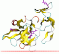
1MYN drosomycin.png 345 × 475; 76 KB
-
Alignment of thioredoxins2.png 435 × 420; 121 KB
-
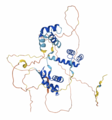
Alpha fold model for DmBCA - Drosophila melanogaster.png 390 × 324; 73 KB
-
Auto-inhibition of Kinesin 1 from Drosophila Melanogaster.gif 700 × 436; 2.04 MB
-
BEN domains of D. melanogaster Insensitive proteins.png 438 × 592; 144 KB
-
Cecropin.png 2,365 × 1,825; 576 KB
-
DptA-Phyre2.jpg 400 × 400; 59 KB
-
Figure 3B (7200354992).png 551 × 592; 420 KB
-
Figure S3A (7784234318).png 706 × 612; 515 KB
-
Figure S4A (7946001558).png 498 × 830; 317 KB
-
Figure S4B (7510918538).png 655 × 719; 686 KB
-
Gooseberry Protein 3D Structure.png 780 × 838; 355 KB
-
Homeodomain-dna-1ahd.png 571 × 644; 179 KB


