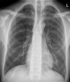
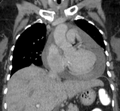
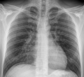
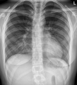
Category:Radiological images by Hellerhoff
Jump to navigation
Jump to search
Deutsch: Diese Kategorie enthält radiologische Bilder aus der Tätigkeit des Benutzers Hellerhoff als Radiologe. Zahlreiche dieser Bilder sind nachbearbeitet (meist in den diagnostisch unwichtigen Bildarealen), um den didaktischen Wert der Bilder zu erhöhen. Die Nutzung der Bilder im Rahmen der jeweils angegebenen Lizenz für Lehrzwecke wird explizit empfohlen. Eine zusätzliche Indizierung der Bilder wurde in dem Projekt pacs.de vorgenommen, wo auch unter hier nicht verwendeten Stichwörtern passende Bilder gefunden werden können. Dort sind auch andere freie radiologische Bilder indiziert, z. T. jedoch nur zur nicht kommerziellen Nutzung (CC nc).
English: This category contains radiological images from the work of the user Hellerhoff as a radiologist. Many of these images have been post-processed (usually in the diagnostically unimportant image areas) to increase the didactic value of the images. The use of the images within the specified license for teaching purposes is explicitly recommended. An additional keywording of the images was carried out in the project pacs.de, where suitable images can also be found under keywords not used here. Other free radiological images are also collected there, some of them only for non-commercial use (CC nc).
Media in category "Radiological images by Hellerhoff"
The following 200 files are in this category, out of 8,813 total.
(previous page) (next page)-
001 Arteriovenous Malformation CT axial 01.png 890 × 1,154; 248 KB
-
001 Arteriovenous Malformation CT axial 02.png 878 × 1,134; 242 KB
-
001 Arteriovenous Malformation CT coronar 01.png 888 × 834; 211 KB
-
001 Arteriovenous Malformation CT coronar 02.png 904 × 828; 200 KB
-
001 Arteriovenous Malformation MRT HAEM axial.png 820 × 1,090; 198 KB
-
001 Arteriovenous Malformation MRT T1KM axial.png 824 × 1,072; 197 KB
-
001 Arteriovenous Malformation MRT T1KM coronar.png 820 × 932; 189 KB
-
001 Arteriovenous Malformation MRT T2 axial.gif 400 × 509; 688 KB
-
001 Arteriovenous Malformation MRT TOF MIP 01.png 1,198 × 1,509; 264 KB
-
001 Arteriovenous Malformation MRT TOF MIP 02.png 1,198 × 1,509; 303 KB
-
001 Arteriovenous Malformation MRT TOF MIP 03.png 1,198 × 1,509; 335 KB
-
001 Arteriovenous Malformation MRT TOF MIP 04.png 1,198 × 1,509; 359 KB
-
001 Arteriovenous Malformation MRT TOF MIP 05.png 1,198 × 1,509; 367 KB
-
001 Arteriovenous Malformation MRT TOF MIP 06.png 1,198 × 1,509; 363 KB
-
001 Arteriovenous Malformation MRT TOF MIP 07.png 1,198 × 1,509; 353 KB
-
001 Arteriovenous Malformation MRT TOF MIP 08.png 1,198 × 1,509; 362 KB
-
001 Arteriovenous Malformation MRT TOF MIP 09.png 1,198 × 1,509; 365 KB
-
001 Arteriovenous Malformation MRT TOF MIP 10.png 1,198 × 1,509; 381 KB
-
001 Arteriovenous Malformation MRT TOF MIP 11.png 1,198 × 1,509; 392 KB
-
001 Arteriovenous Malformation MRT TOF MIP 12.png 1,198 × 1,509; 384 KB
-
001 Arteriovenous Malformation MRT TOF MIP 13.png 1,198 × 1,509; 355 KB
-
001 Arteriovenous Malformation MRT TOF MIP 14.png 1,198 × 1,509; 315 KB
-
001 Arteriovenous Malformation MRT TOF MIP 15.png 1,198 × 1,509; 268 KB
-
001-Arteriovenous-malformation-TOF-MIP-0.jpg 419 × 528; 24 KB
-
001-Arteriovenous-malformation-TOF-MIP-1.jpg 419 × 528; 36 KB
-
001-Arteriovenous-malformation-TOF-MIP-10.jpg 419 × 528; 33 KB
-
001-Arteriovenous-malformation-TOF-MIP-11.jpg 419 × 528; 33 KB
-
001-Arteriovenous-malformation-TOF-MIP-12.jpg 419 × 528; 34 KB
-
001-Arteriovenous-malformation-TOF-MIP-13.jpg 419 × 528; 35 KB
-
001-Arteriovenous-malformation-TOF-MIP-14.jpg 419 × 528; 36 KB
-
001-Arteriovenous-malformation-TOF-MIP-2.jpg 419 × 528; 34 KB
-
001-Arteriovenous-malformation-TOF-MIP-3.jpg 419 × 528; 31 KB
-
001-Arteriovenous-malformation-TOF-MIP-4.jpg 419 × 528; 28 KB
-
001-Arteriovenous-malformation-TOF-MIP-5.jpg 419 × 528; 24 KB
-
001-Arteriovenous-malformation-TOF-MIP-6.jpg 419 × 528; 27 KB
-
001-Arteriovenous-malformation-TOF-MIP-7.jpg 419 × 528; 29 KB
-
001-Arteriovenous-malformation-TOF-MIP-8.jpg 419 × 528; 32 KB
-
001-Arteriovenous-malformation-TOF-MIP-9.jpg 419 × 528; 33 KB
-
01-Sigmadivertikulitis CT ax 001 Perforation - Annotation.png 1,036 × 716; 534 KB
-
01-Sigmadivertikulitis CT ax 001 Perforation.png 1,036 × 716; 510 KB
-
01-Sigmadivertikulitis CT cor 001 Perforation.png 1,134 × 1,082; 693 KB
-
01-Sigmadivertikulitis Sono 001 Verdickte Wand.png 982 × 908; 736 KB
-
01-Sigmadivertikulitis Sono 002 Verdickte Wand.png 840 × 850; 640 KB
-
01-Spondylolyse L5S1 CT axial 001.jpg 1,457 × 954; 114 KB
-
01-Spondylolyse L5S1 CT sagittal 001.jpg 1,031 × 794; 102 KB
-
01-Spondylolyse L5S1 CT sagittal 002.jpg 1,058 × 775; 122 KB
-
02-01-Infiltrat pa.png 1,084 × 1,154; 855 KB
-
02-02-Infiltrat seitlich.png 974 × 1,394; 818 KB
-
02-Sigmadivertikulitis CT ax 001 Umgebung.png 1,104 × 932; 724 KB
-
02-Sigmadivertikulitis CT ax 001 Verdickte Wand.png 1,092 × 834; 656 KB
-
02-Sigmadivertikulitis CT cor 001 Verdickte Wand Umgebungsreaktion.png 1,072 × 1,104; 755 KB
-
03-01-Infiltrat Ausgang.png 884 × 1,066; 546 KB
-
03-02-Infiltrat Verlauf.png 926 × 1,054; 556 KB
-
03-Sigmadivertikulitis CT ax 001 Divertikulose.png 1,046 × 850; 629 KB
-
03-Sigmadivertikulitis CT ax 001 Kleiner Abszess.png 1,050 × 856; 607 KB
-
03-Sigmadivertikulitis CT ax 002 Kleiner Abszess.png 1,042 × 850; 610 KB
-
03-Sigmadivertikulitis CT cor 001 Kleiner Abszess.png 1,026 × 1,012; 566 KB
-
04-01-Infiltrat Ausgang - Verlauf.png 1,946 × 1,006; 1.11 MB
-
04-01-Infiltrat Ausgang.png 968 × 1,006; 526 KB
-
04-02-Infiltrat Verlauf.png 978 × 1,046; 556 KB
-
04-Patellaluxation ap im Roentgen.jpg 673 × 1,088; 128 KB
-
04-Patellaluxation tangential im Roentgen.jpg 961 × 847; 88 KB
-
05-Spontanpneumothorax.jpg 1,060 × 1,227; 133 KB
-
07-01-Hiatushernie pa.png 1,144 × 1,066; 708 KB
-
07-01-Hiatushernie seitlich - Annotation.png 814 × 1,036; 497 KB
-
07-01-Hiatushernie seitlich.png 814 × 1,036; 441 KB
-
08-01-Hiatushernie pa wenig Luft.png 1,050 × 1,028; 615 KB
-
08-02-Hiatushernie CT sagittal - Annotation.png 854 × 1,020; 505 KB
-
08-02-Hiatushernie CT sagittal.png 854 × 1,020; 423 KB
-
08-03-Hiatushernie CT axial.png 1,000 × 852; 461 KB
-
09-01-Pneumothorax Drainage.png 1,050 × 936; 573 KB
-
09-01-Pneumothorax.png 952 × 956; 535 KB
-
10-01-Pneumothorax - Annotation.png 962 × 1,086; 681 KB
-
10-01-Pneumothorax.png 962 × 1,086; 620 KB
-
10-02-Pneumothorax seitlich.png 818 × 1,094; 508 KB
-
10-03-Pneumothorax CT coronar.png 960 × 1,028; 477 KB
-
10-04-Pneumothorax Drainage.png 1,134 × 1,102; 729 KB
-
11-01-Hautfalten Bettlunge - KEIN Pneumothorax.png 1,140 × 1,094; 726 KB
-
12-01-Hautfalten Erguss Bettlunge - KEIN Pneumothorax.png 994 × 1,096; 616 KB
-
14-01-Erguss im Liegen.png 1,140 × 1,078; 655 KB
-
14-02-Erguss im Stehen.png 1,052 × 1,050; 641 KB
-
16-01-Lungenoedem.png 950 × 862; 464 KB
-
16-02-Lungenoedem Verlauf nach 6 Tagen.png 992 × 1,044; 580 KB
-
17-01-Lungenoedem - Ueberwaesserung 19 Jahre nach OP.png 842 × 1,002; 510 KB
-
18-01-Lungenoedem CT coronar.png 1,086 × 1,038; 334 KB
-
20-01-Bettlunge freie Luft Anastomoseninsuffizienz.png 1,006 × 926; 667 KB
-
20-02-Anastomoseninsuffizienz freie Luft - CT.png 1,128 × 1,004; 559 KB
-
20-03-Anastomoseninsuffizienz freie Luft - CT2.png 970 × 868; 452 KB
-
23-04-Achalasie pa nach Dilatation.png 952 × 1,110; 588 KB
-
28-01-Perikarderguss Perimyokarditis pa.png 1,008 × 1,032; 593 KB
-
28-02-Perikarderguss Perimyokarditis CT.png 1,234 × 1,141; 450 KB
-
29-01-Perikarderguss 20 Jahre Perimyokarditis pa Clostridien.png 924 × 1,088; 615 KB
-
29-02-Perikarderguss 20 Jahre Perimyokarditis CT Clostridien.png 1,040 × 678; 287 KB
-
33-01-Pericarditis calcarea pa.png 1,234 × 1,082; 836 KB
-
33-02-Pericarditis calcarea seitlich.png 880 × 1,106; 557 KB
-
34-01-Freie Luft nach LH-OP.png 1,106 × 1,018; 663 KB
-
43-01-Pleuraschwielen Tbc.png 1,088 × 1,112; 729 KB
-
44-01-Nebenbefund Schulter-TEP-Luxation links.png 1,272 × 1,014; 730 KB
-
44-03-Schulter-TEP-Luxation.png 406 × 681; 134 KB
-
44-04-Schulter-TEP nach Reposition.png 768 × 958; 418 KB
-
56-03-Rippenfrakturen - Thorax Pneu 2 Tage spaeter.png 978 × 1,112; 637 KB
-
57-01-Trichterbrust PA.png 1,004 × 1,106; 736 KB
-
57-02-Trichterbrust seitlich.png 720 × 954; 527 KB
-
6-gliedrige LWS 51M - CR - 001 - Annotation.jpg 1,558 × 1,682; 328 KB
-
6-gliedrige LWS 51M - CR - 001.jpg 1,558 × 1,682; 306 KB
-
A lienalis mit erheblichem Gefaesskalk 72M - CR ap - 001.jpg 1,809 × 1,092; 299 KB
-
A lusoria mit erheblichen Verkalkungen 85W - CR pa - 001 - Annotation.jpg 1,714 × 1,228; 224 KB
-
A lusoria mit erheblichen Verkalkungen 85W - CR pa - 001.jpg 1,714 × 1,228; 209 KB
-
A-lusoria CT.jpg 2,292 × 927; 290 KB
-
A-lusoria Schluckuntersuchung.jpg 590 × 696; 80 KB
-
AAI-Schrittmacher 32W - CR pa - 001.jpg 1,479 × 1,914; 214 KB
-
AAI-Schrittmacher bei Sick-Sinus-Syndrom 74M - CR pa - 001.jpg 1,074 × 1,057; 162 KB
-
Abgerissene Drainage im Unterbauch - CT axial.jpg 1,014 × 724; 83 KB
-
Abgerissene Drainage im Unterbauch - CT VR.jpg 570 × 786; 49 KB
-
Abgerissener Drainageschlauch im rechten Abdomen 83W - CR und CT - 001.jpg 2,017 × 1,284; 230 KB
-
Abgerissener Portkatheter und Bergung mit Snare 30W - CR und RF - 001.jpg 1,060 × 1,119; 132 KB
-
Abgerissener Portkatheter und Bergung mit Snare 67W - CR pa und RF - 001.jpg 1,207 × 1,427; 139 KB
-
Ablatio mammae im Roentgenbild 77W - CR pa - 001 - Annotation.jpg 1,086 × 1,111; 189 KB
-
Ablatio mammae im Roentgenbild 77W - CR pa - 001.jpg 1,086 × 1,111; 185 KB
-
Ablatio mammae rechts 46W - CR pa - 001.jpg 1,152 × 1,088; 140 KB
-
Abriss Trochanter major - Krallenplatte mit Cerclagen 78M - CR ap - 001.jpg 1,446 × 918; 205 KB
-
Abrissfraktur des Trochanter major rechts mit Versorgung 83W - CR - 001.jpg 1,951 × 1,314; 279 KB
-
Abscherung am Ursprung des Retinaculum musculorum peroneorum 56W - CR ap - 001.jpg 1,249 × 1,441; 217 KB
-
Abszess in der Milz bei Salmonella O - CT KM - 001 - Annotation.jpg 740 × 562; 68 KB
-
Abszess in der Milz bei Salmonella O - CT KM - 001.jpg 740 × 562; 67 KB
-
AC-Gelenksprengung links in der Panoramaaufnahme 69M - CR ap - 001.jpg 1,532 × 668; 137 KB
-
AC-Gelenkssprengung rechts Roe 001.png 2,824 × 804; 1.35 MB
-
AC-TightRope coracoclavicular intraoperative Durchleuchtung 27W - 001.jpg 2,015 × 918; 112 KB
-
ACG-Sprengung Tossy 3.png 1,534 × 459; 422 KB
-
Achalasie im Breischluck 001.png 612 × 1,545; 427 KB
-
Achalasie im Breischluck 002.png 1,020 × 1,818; 1.2 MB
-
Achalasie im Breischluck 003.png 867 × 1,524; 561 KB
-
Achalasie im Roentgenbild 59W - CR pa - 001.jpg 1,204 × 1,356; 236 KB
-
Achalasie im Roentgenbild 59W - CR seitlich - 001.jpg 1,334 × 1,786; 277 KB
-
Achalasie im Thorax pa 001.png 1,479 × 1,665; 1.2 MB
-
Achalasie im Thorax seitlich 001.png 1,200 × 1,881; 1.03 MB
-
Achalasie in Thoraxübersicht.jpg 2,044 × 2,079; 843 KB
-
Achalasie Stadium 1 mit typischem Tropfen.jpg 633 × 1,108; 57 KB
-
Achalasie Stadium 1.jpg 325 × 850; 58 KB
-
Achalasie Stadium 2.jpg 430 × 837; 68 KB
-
Achalasie Stadium 3 - CT.jpg 877 × 1,158; 197 KB
-
Achalasie Stadium 3 Thorax.jpg 789 × 885; 115 KB
-
Achalasie2-3.jpg 396 × 875; 26 KB
-
Achalasie3.jpg 623 × 874; 49 KB
-
Achillessehnenruptur Sono.jpg 888 × 1,096; 162 KB
-
ACI-Verschluss fehlendes flow-void 70M - MR T2 axial und CT KM - 001.jpg 1,534 × 569; 93 KB
-
Acquired hepatocerebral degeneration MRI T1ax.png 896 × 1,002; 208 KB
-
Acquired hepatocerebral degeneration MRI T1cor.png 752 × 848; 198 KB
-
Acromion Ossifikationszentren.jpg 1,021 × 1,044; 105 KB
-
Actinomyces meyeri 67jm mit Pleuraempyem - CT WF und LF ax - 001.jpg 3,000 × 2,196; 500 KB
-
Actinomyces meyeri 67jm mit Pleuraempyem - Roe pa und CT WF ax - 001.jpg 2,914 × 1,323; 258 KB
-
Adenokarzinom der Gallenblase mit Satellitenherden 66W - CT - 001.jpg 2,028 × 1,428; 290 KB
-
Adenokarzinom der Lunge im Roentgenbild 63M - CR pa - 001.jpg 1,194 × 1,092; 87 KB
-
Adenokarzinom der Lunge im Roentgenbild 63M - CR pa nach Lobektomie - 001.jpg 1,151 × 1,085; 88 KB
-
Adenokarzinom der Lunge im Roentgenbild 63M - CT axial - 001.jpg 1,344 × 1,045; 161 KB
-
Adenokarzinom des Duodenums 65M - CT axial KM pv - 001.jpg 1,097 × 827; 77 KB
-
Adenokarzinom des Duodenums 65M - MR ADC - 001.jpg 1,134 × 794; 57 KB
-
Adenokarzinom des Duodenums 65M - MR DWI - 001.jpg 1,122 × 858; 37 KB
-
Adenokarzinom des Duodenums 65M - MR single shot MRCP - 001.jpg 1,063 × 1,073; 42 KB
-
Adenokarzinom des Duodenums 65M - MR T1 coronar KM - 001.jpg 1,036 × 1,124; 91 KB
-
Adenokarzinom des Duodenums 65M - MR T2 axial - 001.jpg 985 × 731; 59 KB
-
Adenokarzinom des Duodenums 65M - MR T2 FS axial - 001.jpg 1,058 × 844; 48 KB
-
Adenokarzinom des Duodenums 65M - MR T2 FS coronar - 001.jpg 862 × 1,013; 42 KB
-
Adenomyomatose der Gallenblase 71W - MR - 001.jpg 2,009 × 989; 189 KB
-
Aderhautmelanom MRT FLAIR sagittal.jpg 854 × 864; 60 KB
-
Aderhautmelanom MRT T1 axial.jpg 826 × 972; 60 KB
-
Aderhautmelanom MRT T1 KM axial.jpg 1,026 × 808; 68 KB
-
Aderhautmelanom MRT T1 KM koronar.jpg 999 × 970; 67 KB
-
Aderhautmelanom MRT T2 axial.jpg 835 × 962; 82 KB
-
Adulter Granulosazelltumor des Ovars 78jw - CT axial - 001.jpg 1,062 × 1,674; 193 KB
-
Adulter Granulosazelltumor des Ovars 78jw - CT sag - 001.jpg 1,796 × 1,112; 202 KB
-
Aegyptische Fussform 28W - CR ap - 001.jpg 451 × 1,115; 40 KB
-
Aelterer Nierenteilinfarkt links kaudaler Pol 61M - CT coronar KM arteriell - 001.jpg 1,598 × 1,123; 154 KB
-
Aerobilie bei biliodigestiver Anastomose 48M - CT axial KM pv - 001.jpg 1,644 × 1,160; 147 KB
-
Aggregat Hirnstimulator 75jm - Roe pa - 001.jpg 1,122 × 1,117; 103 KB
-
AICA-Loop 69jw - MRT T2 ax - 001.jpg 1,599 × 1,128; 87 KB
-
AICA-Loop 69jw - MRT T2 ax - 002.jpg 1,599 × 1,128; 95 KB
-
Aitken 1 distaler Radius - 12jm - Roe 2Eb - 001.jpg 1,318 × 1,068; 111 KB
-
Aitken I- Salter-Harris- II Fraktur des distalen Radius 9M - CR 2 Ebenen - 001.jpg 1,478 × 1,312; 258 KB
-
Aitken-I-Fraktur der distalen Tibia 11W - CR und CT - 001.jpg 1,840 × 1,348; 292 KB
-
Akin-Osteotomie - Schema - 001.gif 738 × 932; 300 KB
-
Akustikus-Schwannon links intrameatal MRT T1KM axial - Annotation.jpg 1,009 × 840; 81 KB
-
Akustikus-Schwannon links intrameatal MRT T1KM axial.jpg 1,009 × 840; 79 KB
-
Akustikus-Schwannon rechts MRT T1KM axial 001.jpg 956 × 1,085; 80 KB
-
Akustikus-Schwannon rechts MRT T1KM axial 002.jpg 887 × 1,095; 78 KB
-
Akustikus-Schwannon rechts MRT T1KM coronar 001.jpg 880 × 1,015; 80 KB
-
Akustikus-Schwannon rechts MRT T2 axial 001.jpg 889 × 1,027; 100 KB
-
Akute Appendizitis 37W - CT sagittal KM pv - 001.jpg 850 × 1,095; 65 KB
-
Akute Cholezystitis 85M - CT KM pv - 001.jpg 982 × 745; 67 KB
-
Akute exsudative Pankreatitis - CT axial.jpg 1,208 × 1,017; 153 KB
-
Akute Sinusitis maxillaris im MRT 80M - MR T2 axial - 001.jpg 1,123 × 1,399; 122 KB


