Category:Protein-drug interactions
Jump to navigation
Jump to search
Media in category "Protein-drug interactions"
The following 151 files are in this category, out of 151 total.
-
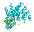
1d2s.jpg 1,000 × 794; 202 KB
-
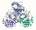
1LG5-cilengitide.png 783 × 617; 152 KB
-
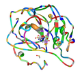
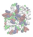
1udt.png 328 × 314; 83 KB
-
2GQG Abl1Kinase Dasatinib.png 1,102 × 847; 555 KB
-
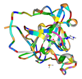
2xp2.png 517 × 403; 159 KB
-
30S streptomycin complex.png 913 × 607; 541 KB
-
3CS9 Abl1 Nilotinib.png 1,071 × 787; 550 KB
-
3D.Sild.2.png 328 × 314; 94 KB
-
3GZJ.png 1,600 × 1,200; 640 KB
-
3LN1.gif 800 × 450; 1.22 MB
-
41375 2020 954 Fig1 HTML.jpg 647 × 603; 74 KB
-
4DAJ.png 795 × 929; 317 KB
-
4ITO.png 370 × 340; 77 KB
-
4ITO2.png 535 × 516; 137 KB
-
5AR contacts.png 1,074 × 436; 253 KB
-
5AR sideview with NADPH and monoolein binding.png 1,038 × 1,204; 694 KB
-
6lu7 wwPDB.png 1,290 × 1,080; 678 KB
-
6wgt 5-HT2A-Rezeptorstruktur.png 608 × 707; 371 KB
-
A general letrozole pharmacophore model.png 1,078 × 948; 116 KB
-
-
Abcwiku.png 497 × 348; 7 KB
-
Actie Site of Eflornithine.png 494 × 515; 223 KB
-
Aldose reductase 1us0.png 1,000 × 878; 499 KB
-
Antivirals.tif 1,920 × 1,080; 1.41 MB
-
Aplaviroc binding.svg 512 × 356; 31 KB
-
Argatroban pocket binding.jpg 207 × 117; 8 KB
-
AryloA2.png 885 × 590; 339 KB
-
AryloCLepB.png 885 × 590; 307 KB
-
Asoprisnil-1.jpg 931 × 927; 71 KB
-
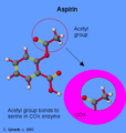
Aspirin blocking COX.png 340 × 357; 10 KB
-
B2appendEps15c.jpg 388 × 390; 37 KB
-
Bafetinib in binding site.PNG 443 × 265; 7 KB
-
Bestatin.jpg 598 × 414; 130 KB
-
Binding interactions between HER2 and Afatinib.svg 833 × 290; 102 KB
-
Binding Mode.jpg 500 × 443; 170 KB
-
Binding of roflumilast to PDE4 enzyme.jpg 521 × 649; 25 KB
-
Binding of SU-6668 to VEGFR-1.svg 196 × 239; 60 KB
-
Binding pockets that aliskiren connects with..png 1,951 × 1,427; 46 KB
-
Bindingsite.jpg 483 × 320; 24 KB
-
Bindisætiogsumatriptan12.PNG 549 × 385; 11 KB
-
Bis(indolyl)maleimide-co-crystallized-catalytic subunit.svg 385 × 387; 87 KB
-
BoroPro in dipeptidyl peptidase IV.png 1,220 × 954; 134 KB
-
Bryettskvma.png 829 × 360; 9 KB
-
Brynja14.png 545 × 348; 7 KB
-
Brynja3.PNG 626 × 440; 12 KB
-
Brynja4.PNG 593 × 353; 11 KB
-
Bygging copy.jpg 241 × 240; 41 KB
-
Cabotegravir Bindung an Integrase.svg 331 × 210; 45 KB
-
Carbose binding to NtMGAM.png 1,134 × 696; 330 KB
-
Citrate Zoom Final.png 640 × 480; 135 KB
-
Complex of Ivacaftor bound to CFTR.png 2,578 × 1,230; 1.63 MB
-
Complex of Ivacaftor with CFTR.png 1,440 × 1,006; 808 KB
-
Cyclopamine in Smoothened Receptor.png 1,229 × 1,008; 225 KB
-
DHFR methotrexate.jpg 767 × 598; 77 KB
-
Docking.jpg 1,272 × 893; 169 KB
-
DOCKING.jpg 633 × 701; 131 KB
-
Dopamine d1 receptor in complex with agonist dopamine.png 1,784 × 1,216; 988 KB
-
Eflornithine in the Active Site.png 325 × 537; 321 KB
-
Efnatengi agonista við amínósýrur.jpg 590 × 481; 38 KB
-
Efnatengi agónista við amínósýrur.jpg 566 × 446; 42 KB
-
Electron Density.jpg 500 × 293; 103 KB
-
Enantiomeric binding sites.png 1,906 × 958; 631 KB
-
Estructura de la P38 unida a un inhibidor.jpg 828 × 518; 88 KB
-
Fig2.Efficacy of Paxlovid against COVID-19.png 600 × 351; 120 KB
-
Figure 2 (7312047000).png 952 × 312; 230 KB
-
Figure 4 (7625036040).png 769 × 695; 632 KB
-
Figure 4. Crystal structure of ERAP1 co-crystallized with DG046..pdf 460 × 452; 113 KB
-
Figure 5 (7069314693).png 843 × 825; 840 KB
-
Figure 8 (6936925035).png 631 × 658; 641 KB
-
Fkbp-cartoon-1fkj.png 730 × 604; 143 KB
-
Fkbp-surface-1fkj.png 764 × 672; 292 KB
-
FKBP25.png 2,000 × 2,000; 580 KB
-
Flottamynd1.png 776 × 360; 9 KB
-
Flottamynd22.png 750 × 316; 9 KB
-
Flottust.PNG 607 × 429; 12 KB
-
Flow chart for structure based drug design.jpg 1,487 × 1,240; 143 KB
-
Flurbiprofen in COX-2.png 605 × 453; 89 KB
-
Fluvastatin bounded to HMG-CoA reductase.png 3,677 × 2,712; 118 KB
-
FXa-rivaroxaban.png 3,000 × 2,034; 2.27 MB
-
GAR transformylase active site with folate based inhibitor.png 1,684 × 983; 581 KB
-
Gefitinib 3d.png 902 × 714; 290 KB
-
GLP-1R complex.png 2,160 × 3,840; 2.06 MB
-
GyraseCiproTop.png 819 × 691; 515 KB
-
HDC Active Site Diagram.tif 960 × 720; 357 KB
-
HGM-coA Reductase with compactin.png 300 × 300; 53 KB
-
HIV protease-indinavir complex.jpg 1,401 × 670; 95 KB
-
HIV1 protease with lipinavir 1MUI 1 sele.png 640 × 480; 56 KB
-
Human CES1 1mx1.jpg 500 × 500; 95 KB
-
IDD594 protein-inhibitor complex.jpg 720 × 540; 54 KB
-
IGFR complexed with allosteric inhibitor (9734919219).jpg 1,280 × 740; 101 KB
-
Imatinib in its binding site.svg 1,646 × 642; 73 KB
-
K252 and c-met.png 337 × 289; 21 KB
-
Liver X Receptor Binding.png 2,731 × 2,319; 1.29 MB
-
LXR-RXR bound polar contacts.png 3,038 × 2,420; 1.62 MB
-
Maraviroc binding.svg 512 × 387; 33 KB
-
Marawirok-bindings.svg 927 × 993; 81 KB
-
MetAP2 bound to fumagillin.png 911 × 538; 460 KB
-
Myndmin.png 724 × 374; 9 KB
-
Médicament.gif 337 × 309; 4 KB
-
Nilotinib in binding site.PNG 387 × 266; 5 KB
-
Nirtalmatrelvir on 3CL.png 475 × 525; 146 KB
-
Nutlin bound to MDM2.png 2,474 × 2,127; 1.06 MB
-
Określanie powinowactwa ligandu do białka.jpg 813 × 353; 39 KB
-
Orthorutheniumbinding pocket2.png 616 × 529; 227 KB
-
PARP1 binding olaparib 5DS3.png 1,440 × 1,207; 1.12 MB
-
Penicillin enzyme complex.jpg 282 × 305; 10 KB
-
Ponatinib in binding site.PNG 356 × 327; 8 KB
-
Ponatinib in its binding site.svg 1,095 × 788; 52 KB
-
Q1 tamoxifen 2.png 2,944 × 1,710; 1.24 MB
-
Quinacrine mustard in Trypanothione reductase active site.png 800 × 582; 292 KB
-
Rassart Figure 1.jpg 1,367 × 474; 126 KB
-
Rifampicin RNApol.png 980 × 595; 241 KB
-
Ritonavir Schechter Berger Notation.svg 356 × 193; 33 KB
-
Rivaroxaban binding pocket sites vizualized.png 2,076 × 928; 77 KB
-
Rivaroxaban Binding To Pockets.jpg 1,152 × 648; 97 KB
-
Roflumilast binding to PDE4 enzyme.jpg 521 × 649; 32 KB
-
Rosuvastatina.jpg 600 × 386; 56 KB
-
Ru-Sav-complex.webm 13 s, 1,190 × 606; 6.46 MB
-
Sarmynd2.jpg 359 × 317; 28 KB
-
Schematic of cocaine ligand structure.png 745 × 370; 94 KB
-
Schild regression bindings.jpg 3,372 × 492; 120 KB
-
Serotoninbinding.jpg 812 × 588; 94 KB
-
Sirolimus binding sites.svg 512 × 476; 60 KB
-
Skjalidokk.png 782 × 374; 9 KB
-
Solbrymaba.png 714 × 374; 9 KB
-
Sorafenib BRAF Labeled.png 1,074 × 927; 701 KB
-
Staphylococcus aureus F98Y DHFR complexed with iclaprim and NADPH.gif 800 × 512; 1.52 MB
-
Structural interaction of rimonabant with CB1.png 738 × 502; 1.42 MB
-
Strúktúrinn.png 776 × 360; 9 KB
-
Substance 14 in its binding site.svg 741 × 350; 30 KB
-
Substrate-like inhibitors and binding to the DPP-4 complex.png 265 × 140; 3 KB
-
Sumatriptan binding site.svg 834 × 740; 29 KB
-
Thymidylate synthase 1HVY.png 757 × 584; 448 KB
-
Tofacitinib,JAK.png 386 × 262; 24 KB
-
Using genomics to identify causes of drug resistance.png 1,155 × 838; 68 KB
-
VEGFR2 bound to axitinib.gif 800 × 512; 1.49 MB
-
View of aliskiren in the binding complex with human renin.png 2,652 × 1,738; 88 KB
-
WEE1 KINASE DOMAIN IN COMPLEX WITH BOSUTINIB.png 3,251 × 2,250; 1.26 MB
-
Wikipic4.png 503 × 261; 46 KB
-
X-Ray diffraction structure of Cisplatin intra-strand GG adducts.png 1,259 × 1,049; 529 KB
-
Xray crystal structure PDB-7si9.png 1,280 × 980; 394 KB


