Category:Petri dishes cultures of Escherichia coli
Jump to navigation
Jump to search
Media in category "Petri dishes cultures of Escherichia coli"
The following 94 files are in this category, out of 94 total.
-
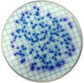
"Петритест®", БГКП, подложка.jpg 1,200 × 952; 273 KB
-
AMG 3724.jpg 3,872 × 2,581; 6.49 MB
-
Antibiogramme.jpg 822 × 856; 552 KB
-
Antibiotic sensitivity and resistance.jpg 2,456 × 1,273; 870 KB
-
Bacteria E.coli.jpg 452 × 604; 30 KB
-
Bacterial culture.jpg 2,500 × 2,091; 2.62 MB
-
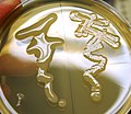
Bacterial smear on nutrient agar - E. coli.JPG 3,648 × 2,736; 1.36 MB
-
Blue-white test.jpg 800 × 800; 288 KB
-
CLED agar after prolonged in incubation.jpg 6,000 × 8,000; 6.79 MB
-
CLED agar.jpg 6,000 × 8,000; 11.4 MB
-
Coli levine.JPG 625 × 464; 42 KB
-
Coliforms.png 1,000 × 1,000; 1.42 MB
-
Colimck.jpeg 728 × 724; 414 KB
-
Colonias de E.Coli en un medio EMB.jpg 2,448 × 3,264; 1.83 MB
-
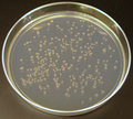
Colonies of Escherichia coli on agar plate.jpg 3,264 × 2,448; 1.4 MB
-
Conjugation.jpg 691 × 518; 55 KB
-
CPSE E.coli.JPG 240 × 320; 27 KB
-
CPSE E.coli2.JPG 640 × 478; 177 KB
-
Differentiation Ecoli Salm.jpg 251 × 240; 24 KB
-
E coli on EMB plate.jpg 3,264 × 2,448; 2.32 MB
-
E coli plate l.jpg 640 × 480; 54 KB
-
E. coli 40X.jpg 2,592 × 1,944; 206 KB
-
E. coli expressing smURFP, a fluorescent protein.tif 1,800 × 800; 2.06 MB
-
E. coli growth on CLED agar.jpg 4,000 × 3,000; 1.2 MB
-
E. coli lactose fermenter (LF) colonies on MacConkey agar.jpg 8,000 × 6,000; 11.47 MB
-
E. coli on EMB agar.png 443 × 452; 260 KB
-
E. coli plate.JPG 706 × 689; 111 KB
-
E. coli sur EMB.jpg 2,322 × 4,128; 2.61 MB
-
E. coli test, bottom.jpg 2,592 × 1,944; 1.11 MB
-
E. coli test, top.jpg 1,944 × 2,592; 1.15 MB
-
E. coli that has been transformed with pGlo under UV light.jpg 4,032 × 3,024; 1.88 MB
-
E.coli JM83 + pUC18.jpg 3,120 × 4,160; 2.06 MB
-
E.coli on growing on various agar media.jpg 2,200 × 1,318; 1.17 MB
-
E.coli on MacConkey agar.JPG 2,048 × 1,360; 1.25 MB
-
E.coli plate dark background.jpg 2,664 × 2,655; 3.15 MB
-
E.coli plate white background.jpg 2,709 × 2,735; 3.75 MB
-
E.coli vs K.pneumoniae.jpg 2,592 × 1,552; 495 KB
-
E.coli-fields.JPG 560 × 560; 125 KB
-
E.coli.Agar levine.jpg 1,024 × 768; 129 KB
-
E.coli.jpg 2,448 × 3,264; 1.69 MB
-
Echerichia coli colony.JPG 2,326 × 1,457; 549 KB
-
Echerichia Coli.jpg 700 × 470; 39 KB
-
Ecoli colonies.png 600 × 542; 427 KB
-
Effectiveness of E.coli.JPG 2,448 × 3,264; 2.05 MB
-
EMB streak plate with E. coli.jpg 1,334 × 1,252; 450 KB
-
Emb-Agar.jpg 616 × 381; 33 KB
-
ESBL Stokes.jpg 2,527 × 2,253; 246 KB
-
Escherichia coli (MCC).jpg 1,373 × 1,371; 932 KB
-
Escherichia coli CCA.png 760 × 748; 1.17 MB
-
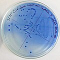
Escherichia coli CLA col.jpg 1,200 × 1,200; 297 KB
-
Escherichia coli colony morphology on CLED medium.jpg 8,000 × 6,000; 11.09 MB
-
Escherichia coli growth on MacConkey agar.jpg 4,000 × 2,250; 3.37 MB
-
Escherichia coli on agar.jpg 2,200 × 1,308; 1.23 MB
-
Escherichia coli, Endo agar.png 705 × 524; 344 KB
-
Eshcherichia coli delta Lon vs wt.jpg 883 × 768; 270 KB
-
Fig1-YhaM-confers-cysteine-resistance.jpg 331 × 336; 18 KB
-
Fluorescent E.coli colonies pGLO.jpg 640 × 480; 54 KB
-
Green colonies of Escherichia coli on agar plate.jpg 3,264 × 2,448; 1.24 MB
-
Green flourescent colonies of Escherichia coli on agar plate.png 2,142 × 1,927; 6 MB
-
Gélose Rambach.JPG 2,272 × 1,704; 1.79 MB
-
KirbyBauer E.coli.JPG 1,047 × 328; 52 KB
-
Lactose fermenting colonies of Escherichia coli.jpg 4,000 × 3,000; 1.34 MB
-
LambdaPlaques.jpg 1,106 × 1,118; 87 KB
-
Love is even in the lab.jpg 720 × 960; 35 KB
-
Macconkey e coli.jpg 610 × 409; 222 KB
-
Mannitol salt agar (MSA) with growth of S. aureus, CoNS and no growth of E. coli.jpg 4,000 × 2,250; 1.88 MB
-
Mannitol Salt Agar with growth of Staphylococcus aureus and CoNS.jpg 4,000 × 2,250; 1.86 MB
-
Modified Hodge Test (MHT).jpg 3,264 × 2,448; 2.12 MB
-
Mucoid strain of Pseudomonas aeruginosa and E. coli growth on CLED agar.jpg 4,000 × 3,000; 1.36 MB
-
National Lab Week 130410-F-TT327-015.jpg 3,600 × 2,395; 908 KB
-
NLF Mucoid colonies of Escherichia coli.jpg 4,000 × 2,250; 1.44 MB
-
Non-lactose fermenting colonies of E. coli on CLED agar.jpg 4,000 × 3,000; 1.13 MB
-
Producentsky kmen.png 549 × 530; 244 KB
-
Producentské kmene.png 2,294 × 2,078; 4.67 MB
-
Salmonellen.e.colli.jpg 4,000 × 3,000; 1.03 MB
-
Staphylococcus, E . coli and Pseudomonas growth on CLED agar.jpg 2,250 × 4,000; 2.57 MB
-
Sterione bild1.png 250 × 260; 152 KB
-
Sterione bild2.png 250 × 258; 157 KB
-
Transformed E.coli using green fluorescent protein 1.jpg 3,000 × 2,000; 935 KB
-
Transformed E.coli using green fluorescent protein 2.jpg 3,000 × 2,000; 765 KB
-
Transformed E.coli using green fluorescent protein 3.jpg 3,000 × 2,000; 810 KB
-
UTI agar.jpg 1,200 × 1,187; 458 KB
-
Voedingsmedium-Ecoli.jpg 1,600 × 1,200; 723 KB
-
XLD Agar-mixed culture of Salmonella sp and Escherichia coli (6500349623).jpg 2,160 × 1,440; 2.11 MB
-
Агар Левина.Escherichia coli.jpg 3,066 × 3,015; 1.27 MB
-
Агар Мак-Конки. Escherichia coli. Лактозопозитивная.jpg 2,978 × 3,111; 893 KB
-
Колонии Кишечной палочки.jpg 6,016 × 4,016; 9.73 MB
-
Колонии Эшерихий.jpg 6,016 × 4,016; 13.36 MB
-
Тризуб-Trident.jpg 4,552 × 3,120; 1.8 MB


