Category:Morphology (biology)
Jump to navigation
Jump to search
study of external forms and structures of organisms | |||||
| Upload media | |||||
| Pronunciation audio | |||||
|---|---|---|---|---|---|
| Instance of | |||||
| Subclass of | |||||
| Part of | |||||
| Facet of |
| ||||
| Has part(s) |
| ||||
| Different from | |||||
| |||||
Subcategories
This category has the following 25 subcategories, out of 25 total.
A
B
C
- Ciliate morphology (11 F)
E
F
H
- Hydrozoa buds (15 F)
I
J
L
M
- Morphologisches Jahrbuch (34 F)
N
- Neuronal morphology (100 F)
P
S
- Synthetic morphology (5 F)
V
Z
Media in category "Morphology (biology)"
The following 74 files are in this category, out of 74 total.
-
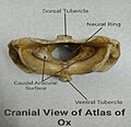
1Cranial-Ox.jpg 514 × 499; 106 KB
-
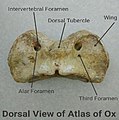
1DorsalOx.jpg 537 × 541; 112 KB
-
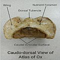
2Caudo-Dorsal-Ox.jpg 497 × 499; 111 KB
-
4Ventral-Ox.jpg 528 × 485; 105 KB
-
5Cranio-Ventral-Ox.jpg 552 × 520; 114 KB
-
Anatomy of turkey head.jpg 450 × 600; 40 KB
-
Aspergillus giganteus morphology.jpg 595 × 795; 100 KB
-
Aurofacial asymmetry.pdf 695 × 385; 155 KB
-
Bird morphology Ashy-crowned Sparrow-lark Male by Dr Raju Kasambe.jpg 3,664 × 2,400; 7.78 MB
-
Black Springbok.svg 1,052 × 744; 58 KB
-
Buttocks morphology.svg 512 × 512; 4 KB
-
De-Morphologie.ogg 1.2 s; 26 KB
-
De-Morphologie2.ogg 2.3 s; 23 KB
-
Different forms of cyanobacteria.webp 3,011 × 1,737; 912 KB
-
Drawing of Loxodes striatus by E Penard.png 762 × 729; 524 KB
-
Drosophila testacea with upward-turned presutural seta in focus (orange arrow).jpg 1,920 × 1,440; 1.4 MB
-
Globorotalia Speciation and Phylogeny.png 535 × 915; 1.87 MB
-
Goldfish male.JPG 1,023 × 1,530; 592 KB
-
Gram Negative Rods of Aeromonas hydrophila.jpg 4,000 × 2,250; 1.08 MB
-
Gram positive cocci in chains of Streptococcus agalactiae.jpg 4,000 × 2,250; 1.4 MB
-
Harpacticoida tagmata.svg 539 × 595; 36 KB
-
Head shot of the species M. niger.png 500 × 312; 194 KB
-
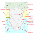
Heteroptera Morphology - English Labels.svg 1,400 × 1,400; 94 KB
-
Jmse-11-00001-g016.png 3,026 × 2,310; 1.59 MB
-
Juanita Vilas Marchant Stenocephalidae Heteroptera HemipteraP.jpg 674 × 960; 419 KB
-
Leioproctus paahaumaa face photograph and entropy image.png 3,191 × 1,361; 4.96 MB
-
Leioproctus paahaumaa face photograph.png 1,653 × 1,361; 2.34 MB
-
Lemna minor 3.jpg 4,320 × 3,240; 5.02 MB
-
Lemna minor 4.jpg 4,320 × 3,240; 5.47 MB
-
Lemna minor 5.jpg 4,320 × 3,240; 5.8 MB
-
Lemna minor fronds 1.jpg 4,320 × 3,240; 5.27 MB
-
Lemna minor fronds 2.jpg 4,320 × 3,240; 5.22 MB
-
Liriodendron tulipifera (Tulip Tree) 14992*A (37501584912).jpg 1,200 × 1,800; 933 KB
-
M. niger Morphology Characteristics.jpg 583 × 469; 53 KB
-
Male facial morphology.jpg 1,184 × 510; 78 KB
-
Mellifera, Sterzeln, Original.jpg 3,859 × 2,573; 6.22 MB
-
Microscopic morphology as a biosignature.jpg 1,330 × 2,712; 929 KB
-
Morfologi bawang merah.jpg 1,477 × 1,108; 77 KB
-
Morfologia de eurypterida.png 717 × 571; 112 KB
-
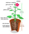
Morfologia vegetal.png 762 × 885; 134 KB
-
Morphological diversity in cyanobacteria.webp 567 × 448; 65 KB
-
Morphological variation within cyanobacterial genera.jpg 3,632 × 2,743; 806 KB
-
Normal Springbok.svg 1,052 × 744; 47 KB
-
Panagrolaimus kolymaensis.png 2,320 × 2,593; 4.83 MB
-
Phenotypic plasticity of colony morphology in Fischerella.webp 473 × 394; 73 KB
-
Potato tuber morphology.svg 751 × 326; 56 KB
-
Schematic representation of Synechocystis cell morphology 2.jpg 1,131 × 843; 180 KB
-
Schematisiertes Ambulacralfüßchen eines Seeigels mit erläuternden Texten.jpg 1,943 × 1,598; 376 KB
-
Selected morphological monographs (1900) (14577354080).jpg 2,262 × 2,938; 531 KB
-
Selected morphological monographs (1900) (14760849991).jpg 2,374 × 2,966; 541 KB
-
Selected morphological monographs (1900) (14760868281).jpg 2,398 × 3,200; 520 KB
-
Selected morphological monographs (1900) (14761669754).jpg 2,282 × 2,928; 502 KB
-
Selected morphological monographs (1900) (14761677804).jpg 2,478 × 3,036; 484 KB
-
Selected morphological monographs (1900) (14761682134).jpg 2,430 × 3,062; 602 KB
-
Selected morphological monographs (1900) (14761690504).jpg 2,410 × 2,976; 505 KB
-
Selected morphological monographs (1900) (14764040865).jpg 2,410 × 2,990; 561 KB
-
Siliquofera grandis subadult female 21.jpg 5,184 × 3,888; 8.47 MB
-
Small to large rod shaped Aeromonas hydrophila in wet mount of culture Microscopy.jpg 4,000 × 2,250; 1.31 MB
-
Superior ovary in Aloe IMG 2029d.jpg 4,032 × 2,898; 2.72 MB
-
Synechocystis cell morphology.webp 3,151 × 1,323; 1.26 MB
-
Tardigrade (unknown species).jpg 1,024 × 943; 79 KB
-
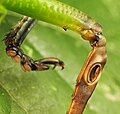
Tympanal Organ of Siliquofera grandis 01.jpg 1,754 × 2,711; 3.86 MB
-
Tympanal Organ of Siliquofera grandis 02.jpg 2,673 × 2,541; 4.77 MB
-
White Springbok.svg 1,052 × 744; 35 KB

