Category:Mikael Häggström/Micrographs of the skin
Jump to navigation
Jump to search
These images were created by Mikael Häggström, M.D.
- User info
- Reusing images
Media in category "Mikael Häggström/Micrographs of the skin"
The following 98 files are in this category, out of 98 total.
-
Compound nevus with moderate atypia.jpg 1,149 × 841; 337 KB
-
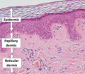
Epidermis, papillary dermis and reticular dermis.png 843 × 737; 1.29 MB
-
Gross pathology of edematous granulation tissue.jpg 484 × 352; 61 KB
-
Gross pathology of squamous cell carcinoma.jpg 1,159 × 861; 229 KB
-
High-magnification micrograph of basal-cell carcinoma.jpg 1,170 × 1,145; 911 KB
-
Histology of a hair follicle at two levels.jpg 2,985 × 1,913; 3.05 MB
-
Histology of a Pacinian corpuscle.jpg 2,048 × 1,532; 1.12 MB
-
Histopathology of a dermal nevus (original).jpg 2,448 × 3,264; 3.26 MB
-
Histopathology of a dermal nevus.jpg 1,487 × 1,103; 852 KB
-
Histopathology of a hypertrophic scar, medium magnification.jpg 1,555 × 1,527; 948 KB
-
Histopathology of a keloid.jpg 2,048 × 1,532; 621 KB
-
Histopathology of a pigmented basal-cell carcinoma.jpg 1,341 × 1,157; 609 KB
-
Histopathology of actinic keratosis with moderate atypia.jpg 1,359 × 1,153; 669 KB
-
Histopathology of actinic keratosis.jpg 2,048 × 1,532; 637 KB
-
Histopathology of chondrodermatitis nodularis chronica helicis.jpg 1,477 × 713; 357 KB
-
Histopathology of chondroid syringoma.jpg 2,048 × 1,532; 876 KB
-
Histopathology of compound nevus.jpg 2,080 × 1,536; 814 KB
-
Histopathology of dermal edema.jpg 1,625 × 1,329; 473 KB
-
Histopathology of dermal nevus on the ear with traumatic epidermal atypia (original).jpg 3,264 × 2,448; 3.04 MB
-
Histopathology of dermal nevus on the ear with traumatic epidermal atypia.jpg 1,359 × 1,185; 805 KB
-
Histopathology of dermal nevus, high magnification (original).jpg 2,448 × 3,264; 3.16 MB
-
Histopathology of dermal nevus, high magnification.jpg 1,359 × 1,685; 1.36 MB
-
Histopathology of dermal nevus, low magnification (original).jpg 2,448 × 3,264; 2.14 MB
-
Histopathology of dermatofibroma.jpg 3,737 × 2,945; 3.02 MB
-
Histopathology of desmoplasia.jpg 2,080 × 1,536; 599 KB
-
Histopathology of edematous granulation tissue, high magnification.jpg 1,246 × 851; 377 KB
-
Histopathology of edematous granulation tissue, low magnification.jpg 1,246 × 372; 176 KB
-
Histopathology of ganglion cyst.jpg 2,080 × 1,536; 723 KB
-
Histopathology of granulomatous folliculitis.jpg 1,390 × 1,017; 598 KB
-
Histopathology of granulomatous inflammation by a ruptured inclusion cyst.jpg 2,080 × 1,536; 848 KB
-
Histopathology of hair follicle periphery.jpg 1,092 × 921; 585 KB
-
Histopathology of invasive melanoma, high magnification.jpg 2,048 × 1,532; 412 KB
-
Histopathology of invasive squamous cell carcinoma.jpg 737 × 565; 247 KB
-
Histopathology of micronodular basal-cell carcinoma (original).jpg 2,448 × 3,264; 2.3 MB
-
Histopathology of micronodular basal-cell carcinoma.jpg 1,085 × 741; 411 KB
-
Histopathology of non-dysplastic dermal nevus, low magnification.jpg 1,359 × 1,039; 576 KB
-
Histopathology of pagetoid dyskeratosis in epidermis of hemorrhoid.jpg 2,048 × 1,532; 1,000 KB
-
Histopathology of pilomatricoma, high magnification, annotated.jpg 2,048 × 1,532; 912 KB
-
Histopathology of pilomatricoma, high magnification.jpg 2,048 × 1,532; 1.06 MB
-
Histopathology of pilomatricoma, low magnification.jpg 2,048 × 1,532; 912 KB
-
Histopathology of pyogenic granuloma - high magnification.jpg 2,048 × 1,532; 664 KB
-
Histopathology of pyogenic granuloma, high magnification, annotated.jpg 2,048 × 1,532; 682 KB
-
Histopathology of radically excised basal-cell carcinoma with separation artifact.jpg 1,358 × 1,627; 778 KB
-
Histopathology of reactive hyperkeratosis.jpg 2,080 × 1,536; 714 KB
-
Histopathology of ruptured epidermoid cyst.jpg 2,048 × 1,532; 619 KB
-
Histopathology of seborrheic keratosis, high magnification (original).jpg 2,448 × 3,264; 2.81 MB
-
Histopathology of seborrheic keratosis, high magnification.jpg 1,023 × 1,294; 691 KB
-
Histopathology of seborrheic keratosis, low magnification (original).jpg 2,448 × 3,264; 2.15 MB
-
Histopathology of seborrheic keratosis, low magnification.jpg 1,941 × 854; 613 KB
-
Histopathology of squamous cell carcinoma in situ.jpg 578 × 458; 171 KB
-
Histopathology of suspected squamous cell carcinoma in re-excision.jpg 587 × 567; 171 KB
-
Histopathology of suture granuloma.jpg 1,149 × 1,059; 687 KB
-
Histopathology of suture material.jpg 2,080 × 1,536; 970 KB
-
Histopathology of trichilemmal cyst - annotated.jpg 1,529 × 1,405; 423 KB
-
Histopathology of trichilemmal cyst.jpg 1,536 × 1,453; 389 KB
-
Low magnification micrograph of a molluscum contagiosum lesion.jpg 2,333 × 1,229; 806 KB
-
Low-level aggressive basal-cell carcinoma.jpg 1,113 × 669; 392 KB
-
Melanophage.jpg 112 × 146; 17 KB
-
Micrograph of a flat wart.jpg 1,697 × 1,659; 929 KB
-
Micrograph of an intradermal melanocytic nevus.jpg 1,281 × 973; 509 KB
-
Micrograph of perinuclear vacuolization (original).jpg 2,448 × 3,264; 2.43 MB
-
Micrograph of perinuclear vacuolization, annotated.jpg 932 × 716; 349 KB
-
Micrograph of squamous cell carcinoma in situ (original).jpg 2,448 × 3,264; 2.37 MB
-
Moderately aggressive basal-cell carcinoma.jpg 601 × 951; 310 KB
-
Nodular basal cell cancer with cleft (original).jpg 2,448 × 3,264; 2.99 MB
-
Nodular basal cell cancer with cleft.jpg 1,047 × 1,015; 493 KB
-
Non-radical basal-cell cancer.jpg 759 × 673; 604 KB
-
Pagetoid peripherally to a melanoma in situ (crop).jpg 623 × 739; 228 KB
-
Pagetoid peripherally to a melanoma in situ.jpg 2,549 × 2,337; 2.1 MB
-
Palisading in basal cell cancer (original).jpg 2,448 × 3,264; 3.56 MB
-
Palisading in basal cell cancer.jpg 1,293 × 1,549; 1.01 MB
-
Pedunculated lipofibroma (intermediate magnification).jpg 1,326 × 1,213; 576 KB
-
Pedunculated lipofibroma (low magnification).jpg 1,326 × 1,704; 366 KB
-
Skin with folds and crush artifact by needle (original).jpg 2,448 × 3,264; 2.87 MB
-
Skin with folds and crush artifact by needle.jpg 1,004 × 803; 406 KB
-
SOX10 immunohistochemistry in a dermal nevus.jpg 681 × 645; 147 KB
-
SOX10 immunohistochemistry of atypical melanocytic proliferation (edited).jpg 1,091 × 446; 175 KB
-
SOX10 immunohistochemistry of atypical melanocytic proliferation (original).jpg 2,448 × 3,264; 1.62 MB
-
SOX10 immunohistochemistry of lentigo maligna (original).jpg 2,448 × 3,264; 1.82 MB
-
SOX10 immunohistochemistry of lentigo maligna.jpg 1,070 × 494; 276 KB
-
SOX10 immunohistochemistry of normal skin (edited).jpg 1,092 × 356; 125 KB
-
SOX10 immunohistochemistry of normal skin (original).jpg 2,448 × 3,264; 1.73 MB
-
Superficial hair follicle tissue.jpg 1,283 × 559; 374 KB
-
Systematic microscopy 1 - Naked eye evaluation, original.jpg 1,185 × 2,527; 694 KB
-
Systematic microscopy 1 - Naked eye evaluation.jpg 2,015 × 997; 529 KB
-
Systematic microscopy 2 - Orientation, original.jpg 4,032 × 3,024; 2.57 MB
-
Systematic microscopy 2 - Orientation.jpg 2,913 × 1,861; 1.75 MB
-
Systematic microscopy 3 - Architectural pattern, original.jpg 2,048 × 1,532; 515 KB
-
Systematic microscopy 3 - Architectural pattern.jpg 1,569 × 1,245; 788 KB
-
Systematic microscopy 4 - Cellular arrangement, original.jpg 2,048 × 1,532; 353 KB
-
Systematic microscopy 4 - Cellular arrangement.jpg 2,048 × 1,532; 545 KB
-
Systematic microscopy 5 - Subcellular features, original.jpg 2,048 × 1,532; 328 KB
-
Systematic microscopy 5 - Subcellular features.jpg 2,048 × 1,532; 539 KB
-
Ulcer border of a squamous cell skin cancer.jpg 1,359 × 1,624; 1.13 MB

