Category:Mikael Häggström/Micrographs of the lung
Jump to navigation
Jump to search
These images were created by Mikael Häggström, M.D.
- User info
- Reusing images
Media in category "Mikael Häggström/Micrographs of the lung"
The following 24 files are in this category, out of 24 total.
-
Histopathology of a pulmonary artery with fat embolism and a bone marrow fragment.jpg 1,765 × 1,233; 770 KB
-
Histopathology of a smoker's macrophage.jpg 549 × 529; 75 KB
-
Histopathology of anthracotic macrophage in lung, annotated.jpg 354 × 349; 48 KB
-
Histopathology of anthracotic macrophage in lung.jpg 354 × 349; 49 KB
-
Histopathology of aspiration.jpg 1,001 × 1,019; 585 KB
-
Histopathology of bronchopneumonia.jpg 1,223 × 1,010; 697 KB
-
Histopathology of chronic pulmonary congestion.jpg 1,109 × 977; 173 KB
-
Histopathology of Gandy–Gamna nodules in chronic pulmonary congestion.jpg 2,048 × 1,532; 356 KB
-
Histopathology of poorly differentiated lung adenocarcinoma.jpg 2,048 × 1,532; 447 KB
-
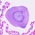
Histopathology of pulmonary alveolar microlithiasis.jpg 1,505 × 1,477; 341 KB
-
Histopathology of pulmonary congestion and siderophages.jpg 969 × 1,094; 254 KB
-
Histopathology of pulmonary hemorrhage in autopsy (zoom).jpg 837 × 651; 259 KB
-
Histopathology of pulmonary hemorrhage in autopsy.jpg 1,739 × 1,484; 1.21 MB
-
Histopathology of respiratory epithelial shedding.jpg 1,111 × 852; 597 KB
-
Histopathology of siderophage in chronic pulmonary congestion.jpg 883 × 833; 115 KB
-
Histopathology of smoker's macrophages with anthracotic stippling.jpg 869 × 727; 175 KB
-
Histopathology of squamous-cell carcinoma of the lung.jpg 1,108 × 818; 345 KB
-
Immunohistochemistry of adenocarcinoma with cytoplasmic staining for TTF-1.jpg 1,353 × 1,037; 294 KB
-
Immunohistochemistry of adenocarcinoma with nuclear staining for TTF-1.jpg 2,048 × 1,532; 252 KB


