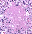
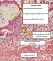
Category:Mikael Häggström/Micrographs of the female reproductive system
Jump to navigation
Jump to search
These images were created by Mikael Häggström, M.D.
- User info
- Reusing images
Media in category "Mikael Häggström/Micrographs of the female reproductive system"
The following 147 files are in this category, out of 147 total.
-
Adequacy counting for cervical Pap tests.png 3,049 × 1,665; 4.74 MB
-
Beta-catenin immunohistochemistry in leiomyoma.jpg 2,048 × 1,532; 343 KB
-
Brenner tumor with annotated coffee bean nuclei.jpg 244 × 250; 35 KB
-
Brenner tumor with microcysts.jpg 1,169 × 1,082; 722 KB
-
Cartilage in a mature cystic teratoma.jpg 1,197 × 1,189; 423 KB
-
Cervical squamous cells with cornflake artifact.jpg 2,048 × 1,532; 769 KB
-
Cytology of cervical squamous metaplasia.png 1,771 × 1,413; 2.58 MB
-
Cytology of endocervical cells.jpg 1,529 × 1,225; 344 KB
-
Cytology of squamous metaplasia.jpg 2,048 × 1,532; 246 KB
-
Cytopathology of keratinizing squamous cell carcinoma.png 965 × 746; 713 KB
-
Cytopathology of low-grade squamous intraepithelial lesion (LSIL).png 1,565 × 981; 1.62 MB
-
Endocervical curetting showing acute cervicitis.jpg 3,500 × 1,536; 914 KB
-
Endometrial endometrioid adenocarcinoma, nuclear and architectural grade 3.jpg 1,072 × 863; 253 KB
-
Epidermis and epidermal appendages a mature cystic teratoma.jpg 1,536 × 2,079; 1.01 MB
-
FIGO grade 2 endometrial adenocarcinoma.jpg 2,048 × 1,532; 797 KB
-
FIGO grade 3 endometrial adenocarcinoma.jpg 2,048 × 1,532; 860 KB
-
Glycogenated cervical squamous cells.tif 2,048 × 1,532; 4.7 MB
-
Gross pathology of a Leydig cell tumor of ovary.jpg 999 × 961; 229 KB
-
Gross pathology of paratubal cysts.jpg 1,237 × 1,057; 322 KB
-
Histology of a Walthard cell rest with coffee bean nuclei.jpg 2,048 × 1,532; 795 KB
-
Histology of a Walthard cell rest, high magnification.jpg 2,048 × 1,532; 647 KB
-
Histology of a Walthard cell rest.jpg 2,048 × 1,532; 779 KB
-
Histology of calcification of a term placenta.jpg 2,048 × 1,532; 495 KB
-
Histology of ciliated columnar epithelium of the fallopian tube.jpg 1,219 × 783; 185 KB
-
Histology of decidua.jpg 2,080 × 1,536; 728 KB
-
Histology of endocervix.jpg 1,313 × 2,020; 810 KB
-
Histology of implantation site intermediate trophoblasts.jpg 2,080 × 1,536; 745 KB
-
Histology of mesonephric duct remnant.jpg 3,074 × 2,048; 2.45 MB
-
Histology of nabothian cysts on cervical biopsy.jpg 2,048 × 1,532; 584 KB
-
Histology of normal simple columnar epithelium of the endometrium.jpg 1,069 × 741; 243 KB
-
Histology of ovarian stroma.jpg 2,048 × 1,532; 597 KB
-
Histology of ovarian surface epithelium.jpg 2,048 × 1,532; 371 KB
-
Histology of postmenopausal myometrium, intermediate magnification.jpg 2,080 × 1,536; 1.02 MB
-
Histology of postmenopausal myometrium, low magnification.jpg 2,080 × 1,536; 1.27 MB
-
Histology of scantly cellular decidua.jpg 2,080 × 1,536; 1,022 KB
-
Histomathology of meconium histocytosis (original).jpg 2,048 × 1,532; 448 KB
-
Histomathology of meconium histocytosis.jpg 2,037 × 1,527; 567 KB
-
Histopathology of a corpus luteum cyst, high magnification.jpg 2,048 × 1,532; 1.03 MB
-
Histopathology of a corpus luteum cyst, low magnification.jpg 2,048 × 1,532; 1.15 MB
-
Histopathology of a leiomyoma in a postmenopausal uterus, intermediate magnification.jpg 2,080 × 1,536; 904 KB
-
Histopathology of a leiomyoma in a postmenopausal uterus, low magnification.jpg 2,080 × 1,536; 998 KB
-
Histopathology of a leiomyoma with fascicular growth.jpg 2,048 × 1,532; 868 KB
-
Histopathology of acute choriodeciduitis (original).jpg 2,048 × 1,532; 483 KB
-
Histopathology of acute choriodeciduitis.jpg 813 × 873; 213 KB
-
Histopathology of adenomyosis.jpg 1,358 × 1,158; 718 KB
-
Histopathology of amnionitis, annotated.jpg 1,195 × 771; 222 KB
-
Histopathology of amnionitis.jpg 2,048 × 1,532; 438 KB
-
Histopathology of an adenomatoid tumor of the fallopian tube, low magnification.jpg 2,121 × 2,045; 1.99 MB
-
Histopathology of an ectopic pregnancy in a fallopian tube.jpg 2,048 × 1,532; 911 KB
-
Histopathology of bony tissue in a mature teratoma.jpg 2,048 × 1,532; 445 KB
-
Histopathology of brain tissue in a mature teratoma.jpg 2,048 × 1,532; 914 KB
-
Histopathology of carcinosarcoma with chondroid matrix.jpg 2,048 × 1,532; 354 KB
-
Histopathology of carcinosarcoma, annotated.png 2,048 × 1,532; 7.13 MB
-
Histopathology of carcinosarcoma, low magnification.jpg 2,048 × 1,532; 685 KB
-
Histopathology of carcinosarcoma.jpg 2,048 × 1,532; 746 KB
-
Histopathology of chorangiosis.jpg 2,080 × 1,536; 764 KB
-
Histopathology of chorioamnionitis.jpg 1,187 × 893; 254 KB
-
Histopathology of chorionic villi at gestational age of 9 weeks.jpg 2,080 × 1,536; 1.05 MB
-
Histopathology of choroid plexus tissue in a mature teratoma.jpg 2,048 × 1,532; 800 KB
-
Histopathology of CIN 3 (original).jpg 2,439 × 2,517; 1.94 MB
-
Histopathology of CIN 3 with endocervical gland invasion (original).jpg 2,448 × 3,264; 3 MB
-
Histopathology of CIN 3 with endocervical gland invasion.jpg 1,148 × 1,200; 747 KB
-
Histopathology of CIN 3 with small endocervical gland invasion.jpg 1,358 × 1,711; 1,004 KB
-
Histopathology of CIN 3.jpg 1,010 × 677; 410 KB
-
Histopathology of edematous fallopian tubes.jpg 2,048 × 1,532; 911 KB
-
Histopathology of endometrial adenocarcinoma, endometrioid type.jpg 2,048 × 1,532; 759 KB
-
Histopathology of endometrial cancer with lymphovascular invasion.jpg 2,048 × 1,532; 1.34 MB
-
Histopathology of endometrial intraepithelial neoplasia (EIN).jpg 2,048 × 1,532; 873 KB
-
Histopathology of endometrioid cancer, grade 1, nuclear grade 1.jpg 1,177 × 969; 406 KB
-
Histopathology of endometrioid cancer, grade 1, nuclear grade 2.jpg 1,107 × 939; 422 KB
-
Histopathology of fascicular growth in a leiomyoma.jpg 2,048 × 1,532; 873 KB
-
Histopathology of fatty tissue in a mature teratoma.jpg 2,048 × 1,532; 551 KB
-
Histopathology of fetal membrane edema as a sign of meconium, annotated.jpg 2,080 × 1,536; 642 KB
-
Histopathology of fetal membrane edema as a sign of meconium.jpg 2,080 × 1,536; 649 KB
-
Histopathology of fibrinoid necrosis of the placenta.jpg 1,221 × 1,285; 432 KB
-
Histopathology of FIGO (architectural) grade 1 endometrial adenocarcinoma.png 1,393 × 1,281; 3.05 MB
-
Histopathology of follicular cervicitis, high magnification.jpg 2,048 × 1,532; 562 KB
-
Histopathology of follicular cervicitis, low magnification.jpg 2,048 × 1,532; 569 KB
-
Histopathology of follicular cervicitis.png 2,897 × 2,173; 9.15 MB
-
Histopathology of grade 2 endometrioid endometrial cancer with myometrial invasion.jpg 1,533 × 1,201; 635 KB
-
Histopathology of high-grade squamous intraepithelial lesion (HSIL) of vulva.jpg 2,048 × 1,532; 860 KB
-
Histopathology of invasive low-grade serous carcinoma of ovary (original).jpg 3,205 × 2,097; 1.47 MB
-
Histopathology of invasive low-grade serous carcinoma of ovary.png 3,209 × 2,189; 9 MB
-
Histopathology of leiomyoma with nuclear pleomorphism.jpg 2,048 × 1,532; 428 KB
-
Histopathology of leydig cell tumor of the ovary, high mag, annotated.png 2,048 × 1,532; 7.05 MB
-
Histopathology of leydig cell tumor of the ovary, high mag.jpg 2,048 × 1,532; 457 KB
-
Histopathology of leydig cell tumor of the ovary, low mag.jpg 2,048 × 1,532; 610 KB
-
Histopathology of moderate fibrinoid necrosis of the placenta.jpg 2,080 × 1,536; 1.11 MB
-
Histopathology of necrotic chorionic villi.jpg 1,797 × 1,193; 789 KB
-
Histopathology of non-complex endometrial polyp without atypia (original).jpg 2,448 × 3,264; 3.41 MB
-
Histopathology of non-complex endometrial polyp without atypia, with tubal metaplasia.jpg 1,814 × 1,195; 1.06 MB
-
Histopathology of non-complex endometrial polyp without atypia.jpg 1,328 × 1,262; 997 KB
-
Histopathology of ovarian serous borderline tumor with non-invasive invaginations.jpg 2,048 × 1,532; 994 KB
-
Histopathology of ovarian serous borderline tumor.jpg 2,867 × 1,977; 1.5 MB
-
Histopathology of paratubal cyst.jpg 453 × 381; 48 KB
-
Histopathology of phlebitis and funisitis, annotated.jpg 962 × 720; 224 KB
-
Histopathology of placenta with increased syncytial knotting of chorionic villi.jpg 2,048 × 1,532; 813 KB
-
Histopathology of Serous carcinoma arising in endometrial polyp.jpg 2,301 × 2,029; 1.52 MB
-
Histopathology of serous carcinoma of uterus.jpg 2,048 × 1,532; 1.01 MB
-
Histopathology of simple squamous cyst wall.jpg 1,310 × 581; 337 KB
-
Histopathology of subchorionic intervillositis, annotated.jpg 1,443 × 1,121; 426 KB
-
Histopathology of subchorionic intervillositis, original.jpg 1,443 × 1,121; 382 KB
-
Histopathology of tubal pregnancy (hy).png 1,267 × 1,462; 2.77 MB
-
Histopathology of tubal pregnancy - original.jpg 2,448 × 3,264; 2.97 MB
-
Histopathology of tubal pregnancy.jpg 1,267 × 1,462; 825 KB
-
Histopathology of tubal pregnancy.svg 950 × 1,097; 435 KB
-
Histopathology of uterine leiomyoma (original).jpg 2,448 × 3,264; 2.63 MB
-
Histopathology of uterine leiomyoma (Van Gieson's stain).jpg 1,107 × 875; 612 KB
-
Histopathology of uterine leiomyoma (Van Gieson's stain, original).jpg 2,448 × 3,264; 3.02 MB
-
Histopathology of uterine leiomyoma.jpg 1,107 × 1,026; 564 KB
-
Histopathology of woven or storiform pattern.jpg 1,877 × 1,265; 934 KB
-
Immunohistochemistry for p16 in cervical squamous cell carcinoma.jpg 1,532 × 2,048; 666 KB
-
Immunohistochemistry of loss of expression of MLH-1.jpg 2,048 × 1,532; 518 KB
-
Immunohistochemistry of retained expression of MSH-6.jpg 2,048 × 1,532; 607 KB
-
Making a fetal membrane roll.jpg 3,029 × 2,337; 1.96 MB
-
Micrograph of a clue cell, annotated.jpg 2,048 × 1,532; 436 KB
-
Micrograph of a clue cell.jpg 2,048 × 1,532; 518 KB
-
Myometrium versus endometrial stroma versus endometrial polyp stroma - Original.jpg 2,048 × 1,532; 1,000 KB
-
Myometrium versus endometrial stroma versus endometrial polyp stroma.jpg 2,048 × 1,532; 772 KB
-
Nervous tissue in a mature cystic teratoma.jpg 2,080 × 1,536; 858 KB
-
Normal vaginal flora on Pap stain.jpg 2,453 × 2,033; 698 KB
-
Normal vaginal flora versus bacterial vaginosis on Pap stain.jpg 5,121 × 4,253; 2.43 MB
-
Respiratory epithelium in a mature cystic teratoma.jpg 1,467 × 713; 201 KB
-
Uterine leiomyoma with lining (original).jpg 2,448 × 3,264; 3.19 MB
-
Uterine leiomyoma with lining.jpg 1,206 × 1,284; 857 KB
-
Vaginal wet mount of candidal vulvovaginitis - original.jpg 907 × 923; 189 KB
-
Vaginal wet mount of candidal vulvovaginitis.jpg 907 × 923; 300 KB
-
Vaginal wet mount with clue cell - annotated.png 809 × 611; 693 KB
-
Woven pattern.jpg 2,481 × 3,377; 3 MB


