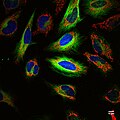
Category:Microscopic images with microtubule specific stains
Jump to navigation
Jump to search
Media in category "Microscopic images with microtubule specific stains"
The following 24 files are in this category, out of 24 total.
-
Birdie's nest.jpg 1,024 × 1,024; 823 KB
-
Btub.jpg 779 × 695; 252 KB
-
Cell divisions in Arabidopsis primary root meristem cells.tif 1,527 × 513; 765 KB
-
Christmas spirit.jpg 408 × 408; 48 KB
-
Cultured Rat Hippocampal Neuron (24327909026).jpg 1,918 × 1,219; 784 KB
-
Different mitotic stages in Arabidopsis primary root meristem cells.tif 774 × 256; 287 KB
-
Dividing Cell Fluorescence-ru.jpg 554 × 554; 165 KB
-
Dividing Cell Fluorescence.jpg 554 × 554; 58 KB
-
Fluorescent image fibroblast.jpg 1,500 × 2,250; 440 KB
-
FluorescentCells.jpg 512 × 512; 56 KB
-
Fox stealing eggs.jpg 693 × 588; 95 KB
-
HeLa-I.jpg 2,400 × 1,999; 2.15 MB
-
HeLa-II.jpg 3,200 × 2,400; 2.76 MB
-
HeLa-Tubulin-HSP60-Fibrillarin-DNA.jpg 2,968 × 2,976; 4.93 MB
-
Hippocampal neurons in culture.tif 1,024 × 1,024; 3.03 MB
-
Ki67-Tubulin-2.jpg 1,156 × 874; 542 KB
-
Ki67-Tubulin.jpg 1,186 × 870; 743 KB
-
Mice embryonic fibroblasts GFP.tif 2,048 × 2,048; 12.01 MB
-
Microtubules in the leading edge of a cell.tif 3,959 × 5,352; 60.65 MB
-
Multicolor fluorescence image of a living HeLa cell.jpg 1,024 × 1,024; 148 KB
-
Multicolor fluorescence image of a living PC-12 cell.jpeg 1,024 × 1,024; 79 KB
-
Multicolor fluorescence image of living HeLa cells.jpg 1,024 × 1,024; 184 KB
-
The internal structure of a migrating cancer cell.jpg 1,024 × 674; 70 KB
-
Two legged goat.jpg 968 × 862; 106 KB

