Category:Microscopic images of insect legs
Jump to navigation
Jump to search
Media in category "Microscopic images of insect legs"
The following 54 files are in this category, out of 54 total.
-
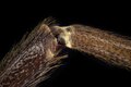
 Acalles camelus metatarsus.jpg 644 × 1,017; 221 KB
-
 Ant leg (250 09) Ant leg (Formicidae).jpg 3,751 × 2,401; 2.48 MB
-
 Autres griffes d' Arachnocoris karukerae.jpg 1,501 × 1,560; 348 KB
-
 Beetle's claws on scanning electron microscope.jpg 895 × 897; 498 KB
-
 Cranefly leg.png 1,280 × 720; 377 KB
-
 Diptera (YPM IZ 098032).jpeg 1,920 × 1,426; 151 KB
-
 Diptera (YPM IZ 098561).jpeg 1,920 × 1,421; 295 KB
-
 Drosophila suzukii sex combs b.jpg 5,556 × 5,556; 6.46 MB
-
 Dysmicoccus brevipes leg translucent pore.jpg 720 × 540; 58 KB
-
 Epines d' Arachnocoris karukerae.jpg 1,010 × 1,006; 196 KB
-
 Epines d' Arachnocoris thesauri.jpg 1,434 × 1,209; 305 KB
-
 Epines d' Arachnocoris varius.jpg 1,444 × 1,232; 253 KB
-
 Fliegenbein 25x.tif 1,024 × 768; 781 KB
-
 Fruit Fly 1.jpg 1,024 × 1,083; 572 KB
-
 Grasshopper Leg.jpg 5,334 × 2,789; 2.24 MB
-
 Griffes d' Arachnocoris karukerae.jpg 1,048 × 1,100; 232 KB
-
 Griffes d' Arachnocoris varius.jpg 2,204 × 1,522; 453 KB
-
 Insect Legs.jpg 5,184 × 3,456; 6.97 MB
-
 JPGR-Beetle EM claw.tif 1,024 × 1,024; 1 MB
-
 MantisLegW40x4.jpg 1,024 × 768; 216 KB
-
 Moth2leg1.jpg 1,278 × 704; 143 KB
-
 Musca coxa micrograph.jpg 1,280 × 1,100; 199 KB
-
 Musca joint closeup micrograph.jpg 1,280 × 1,100; 161 KB
-
 Musca leg disarticulation micrograph.jpg 1,280 × 1,100; 177 KB
-
 Musca leg micrograph.jpg 1,280 × 1,100; 188 KB
-
 Musca pulvilli closeup micrograph.jpg 1,280 × 1,100; 166 KB
-
 Musca pulvilli micrograph.jpg 1,280 × 1,100; 172 KB
-
 Musca tarsus micrograph.jpg 1,280 × 1,100; 177 KB
-
 Pata de cucaracha vista bajo el microscopio electrónico de barrido.jpg 2,048 × 1,392; 1.5 MB
-
 Pseudococcus jackbeardsleyi eye.jpg 720 × 540; 31 KB
-
 Pseudococcus jackbeardsleyi leg - translucent pore.jpg 720 × 540; 56 KB
-
 Stiltleg2.jpg 339 × 639; 38 KB
-
 Stinde Mikrofoto1.JPG 207 × 241; 56 KB
-
 Tarsus of Chrysomelidae.jpg 1,560 × 1,254; 452 KB
-
 TarsusREM.jpg 2,576 × 2,086; 753 KB
-
 Thing.jpg 2,250 × 3,000; 1.01 MB
-
 Trigonopterus KA1 metatarsus.jpg 669 × 1,022; 304 KB
-
 Vespula vulgaris SEM Foreleg 1st Joint.jpg 1,280 × 1,024; 721 KB
-
 Vespula vulgaris SEM Foreleg 2nd Joint with Tibial Spur 01.jpg 1,280 × 1,024; 801 KB
-
 Vespula vulgaris SEM Foreleg 2nd Joint with Tibial Spur 02.jpg 1,280 × 1,024; 789 KB
-
 Vespula vulgaris SEM Legs.jpg 1,280 × 1,024; 702 KB
-
 Лапа мухи львинки в сканирующем электронном микроскопе.jpg 2,302 × 1,332; 4.76 MB
-
 Лапка большого комара долгоножки 2.tif 4,343 × 2,896; 71.99 MB
-
 Лапка бражника.jpg 3,300 × 2,193; 1.85 MB
-
 Лапка комара долгоножки.tif 4,400 × 2,934; 73.89 MB
-
 Лапка кузнечика 1.jpg 5,511 × 9,799; 11.78 MB
-
 Лапка кузнечика 2.jpg 6,583 × 11,702; 16.59 MB
-
 Лапка мухи с нано подушечками.jpg 1,535 × 1,803; 1.72 MB
-
 Лапка мухи.jpg 1,600 × 1,200; 509 KB
-
 Лапки пчелы под микроскопом.jpg 930 × 930; 176 KB
-
 Присоска на лапе жука плавунца.jpg 4,592 × 4,757; 10.56 MB
-
 Присоски на ламе жука плавунца.jpg 4,274 × 3,209; 9.18 MB
-
 Сустав ноги долгоносика в электронном микроскопе.jpg 1,536 × 1,521; 2.28 MB
-
 Сустав ноги муравья в электронном микроскопе.jpg 1,770 × 1,611; 4.5 MB

_Ant_leg_(Formicidae).jpg)


.jpeg)
.jpeg)


